2CJS

Structural Basis for a Munc13-1 Homodimer - Munc13-1 - RIM Heterodimer Switch: C2-domains as Versatile Protein-Protein Interaction Modules


Experimental Data Snapshot

  • Method: X-RAY DIFFRACTION
  • Resolution: 1.78 Å
  • R-Value Free: 0.219 
  • R-Value Work: 0.175 
  • R-Value Observed: 0.176 

wwPDB Validation   3D Report Full Report


This is version 1.3 of the entry. See complete history


Literature

Structural Basis for a Munc13-1 Homodimer to Munc13-1/Rim Heterodimer Switch.

Lu, J.Machius, M.Dulubova, I.Dai, H.Sudhof, T.C.Tomchick, D.R.Rizo, J.

(2006) PLoS Biol 4: E192

  • DOI: https://doi.org/10.1371/journal.pbio.0040192
  • Primary Citation of Related Structures:  
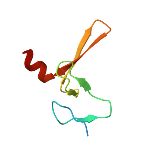
    2CJS, 2CJT

  • PubMed Abstract: 

    C(2) domains are well characterized as Ca(2+)/phospholipid-binding modules, but little is known about how they mediate protein-protein interactions. In neurons, a Munc13-1 C(2)A-domain/RIM zinc-finger domain (ZF) heterodimer couples synaptic vesicle priming to presynaptic plasticity. We now show that the Munc13-1 C(2)A domain homodimerizes, and that homodimerization competes with Munc13-1/RIM heterodimerization. X-ray diffraction studies guided by nuclear magnetic resonance (NMR) experiments reveal the crystal structures of the Munc13-1 C(2)A-domain homodimer and the Munc13-1 C(2)A-domain/RIM ZF heterodimer at 1.44 A and 1.78 A resolution, respectively. The C(2)A domain adopts a beta-sandwich structure with a four-stranded concave side that mediates homodimerization, leading to the formation of an eight-stranded beta-barrel. In contrast, heterodimerization involves the bottom tip of the C(2)A-domain beta-sandwich and a C-terminal alpha-helical extension, which wrap around the RIM ZF domain. Our results describe the structural basis for a Munc13-1 homodimer-Munc13-1/RIM heterodimer switch that may be crucial for vesicle priming and presynaptic plasticity, uncovering at the same time an unexpected versatility of C(2) domains as protein-protein interaction modules, and illustrating the power of combining NMR spectroscopy and X-ray crystallography to study protein complexes.


  • Organizational Affiliation

    Department of Biochemistry, University of Texas Southwestern Medical Center, Dallas, Texas, USA.


Macromolecules
Find similar proteins by:  (by identity cutoff)  |  3D Structure
Entity ID: 1
MoleculeChains Sequence LengthOrganismDetailsImage
UNC-13 HOMOLOG A
A, B
167Rattus norvegicusMutation(s): 1 
Membrane Entity: Yes 
UniProt
Find proteins for Q62768 (Rattus norvegicus)
Explore Q62768 
Go to UniProtKB:  Q62768
Entity Groups  
Sequence Clusters30% Identity50% Identity70% Identity90% Identity95% Identity100% Identity
UniProt GroupQ62768
Sequence Annotations
Expand
  • Reference Sequence
Find similar proteins by:  (by identity cutoff)  |  3D Structure
Entity ID: 2
MoleculeChains Sequence LengthOrganismDetailsImage
REGULATING SYNAPTIC MEMBRANE EXOCYTOSIS PROTEIN 262Rattus norvegicusMutation(s): 0 
Membrane Entity: Yes 
UniProt
Find proteins for Q9JIS1 (Rattus norvegicus)
Explore Q9JIS1 
Go to UniProtKB:  Q9JIS1
Entity Groups  
Sequence Clusters30% Identity50% Identity70% Identity90% Identity95% Identity100% Identity
UniProt GroupQ9JIS1
Sequence Annotations
Expand
  • Reference Sequence
Small Molecules
Ligands 3 Unique
IDChains Name / Formula / InChI Key2D Diagram3D Interactions
GOL
Query on GOL

Download Ideal Coordinates CCD File 
F [auth A]
G [auth A]
H [auth A]
J [auth B]
K [auth B]
F [auth A],
G [auth A],
H [auth A],
J [auth B],
K [auth B],
P [auth C]
GLYCEROL
C3 H8 O3
PEDCQBHIVMGVHV-UHFFFAOYSA-N
ZN
Query on ZN

Download Ideal Coordinates CCD File 
L [auth C],
M [auth C]
ZINC ION
Zn
PTFCDOFLOPIGGS-UHFFFAOYSA-N
EDO
Query on EDO

Download Ideal Coordinates CCD File 
D [auth A],
E [auth A],
I [auth B],
N [auth C],
O [auth C]
1,2-ETHANEDIOL
C2 H6 O2
LYCAIKOWRPUZTN-UHFFFAOYSA-N
Experimental Data & Validation

Experimental Data

  • Method: X-RAY DIFFRACTION
  • Resolution: 1.78 Å
  • R-Value Free: 0.219 
  • R-Value Work: 0.175 
  • R-Value Observed: 0.176 
  • Space Group: P 21 21 21
Unit Cell:
Length ( Å )Angle ( ˚ )
a = 50.248α = 90
b = 93.534β = 90
c = 113.128γ = 90
Software Package:
Software NamePurpose
REFMACrefinement
HKL-2000data reduction
HKL-2000data scaling
PHASERphasing

Structure Validation

View Full Validation Report


Entry History 

Deposition Data

Revision History  (Full details and data files)

  • Version 1.0: 2006-06-07
    Type: Initial release
  • Version 1.1: 2011-07-13
    Changes: Advisory, Version format compliance
  • Version 1.2: 2013-01-30
    Changes: Database references, Derived calculations, Non-polymer description, Structure summary
  • Version 1.3: 2019-01-30
    Changes: Data collection, Experimental preparation, Other