2CIW

Chloroperoxidase iodide complex


Experimental Data Snapshot

  • Method: X-RAY DIFFRACTION
  • Resolution: 1.15 Å
  • R-Value Free: 0.171 
  • R-Value Work: 0.149 
  • R-Value Observed: 0.150 

wwPDB Validation   3D Report Full Report


Ligand Structure Quality Assessment 


This is version 3.1 of the entry. See complete history


Literature

Crystal Structures of Chloroperoxidase with its Bound Substrates and Complexed with Formate, Acetate, and Nitrate.

Kuhnel, K.Blankenfeldt, W.Terner, J.Schlichting, I.

(2006) J Biol Chem 281: 23990

  • DOI: https://doi.org/10.1074/jbc.M603166200
  • Primary Citation of Related Structures:  
    2CIV, 2CIW, 2CIX, 2CIY, 2CIZ, 2CJ0, 2CJ1, 2CJ2

  • PubMed Abstract: 

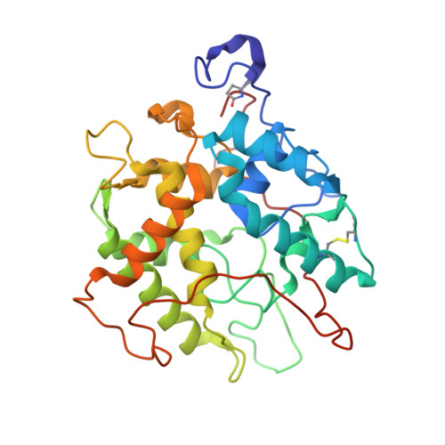
    Chloroperoxidase (CPO) is a heme-thiolate enzyme that catalyzes hydrogen peroxide-dependent halogenation reactions. Structural data on substrate binding have not been available so far. CPO was therefore crystallized in the presence of iodide or bromide. One halide binding site was identified at the surface near a narrow channel that connects the surface with the heme. Two other halide binding sites were identified within and at the other end of this channel. Together, these sites suggest a pathway for access of halide anions to the active site. The structure of CPO complexed with its natural substrate cyclopentanedione was determined at a resolution of 1.8 A. This is the first example of a CPO structure with a bound organic substrate. In addition, structures of CPO bound with nitrate, acetate, and formate and of a ternary complex with dimethylsulfoxide (Me2SO) and cyanide were determined. These structures have implications for the mechanism of compound I formation. Before binding to the heme, the incoming hydrogen peroxide first interacts with Glu-183. The deprotonated Glu-183 abstracts a proton from hydrogen peroxide. The hydroperoxo-anion then binds at the heme, yielding compound 0. Glu-183 protonates the distal oxygen of compound 0, water is released, and compound I is formed.


  • Organizational Affiliation

    Abteilung Biomolekulare Mechanismen, Max-Planck-Institut für Medizinische Forschung, Jahnstrasse 29, 69120 Heidelberg, Germany.


Macromolecules
Find similar proteins by:  (by identity cutoff)  |  3D Structure
Entity ID: 1
MoleculeChains Sequence LengthOrganismDetailsImage
CHLOROPEROXIDASE299Leptoxyphium fumagoMutation(s): 0 
EC: 1.11.1.10
UniProt
Find proteins for P04963 (Leptoxyphium fumago)
Explore P04963 
Go to UniProtKB:  P04963
Entity Groups  
Sequence Clusters30% Identity50% Identity70% Identity90% Identity95% Identity100% Identity
UniProt GroupP04963
Sequence Annotations
Expand
  • Reference Sequence
Oligosaccharides

Help

Entity ID: 2
MoleculeChains Length2D Diagram Glycosylation3D Interactions
beta-D-mannopyranose-(1-4)-2-acetamido-2-deoxy-beta-D-glucopyranose-(1-4)-2-acetamido-2-deoxy-beta-D-glucopyranose
B
3N-Glycosylation
Glycosylation Resources
GlyTouCan:  G15407YE
GlyCosmos:  G15407YE
GlyGen:  G15407YE
Entity ID: 3
MoleculeChains Length2D Diagram Glycosylation3D Interactions
alpha-D-mannopyranose-(1-2)-alpha-D-mannopyranose
C
2O-Glycosylation
Glycosylation Resources
GlyTouCan:  G53402KW
GlyCosmos:  G53402KW
GlyGen:  G53402KW
Small Molecules
Ligands 5 Unique
IDChains Name / Formula / InChI Key2D Diagram3D Interactions
HEM
Query on HEM

Download Ideal Coordinates CCD File 
E [auth A]PROTOPORPHYRIN IX CONTAINING FE
C34 H32 Fe N4 O4
KABFMIBPWCXCRK-RGGAHWMASA-L
NAG
Query on NAG

Download Ideal Coordinates CCD File 
F [auth A],
G [auth A]
2-acetamido-2-deoxy-beta-D-glucopyranose
C8 H15 N O6
OVRNDRQMDRJTHS-FMDGEEDCSA-N
MAN
Query on MAN

Download Ideal Coordinates CCD File 
H [auth A]
I [auth A]
J [auth A]
K [auth A]
L [auth A]
H [auth A],
I [auth A],
J [auth A],
K [auth A],
L [auth A],
M [auth A],
N [auth A],
O [auth A],
P [auth A],
Q [auth A],
R [auth A]
alpha-D-mannopyranose
C6 H12 O6
WQZGKKKJIJFFOK-PQMKYFCFSA-N
IOD
Query on IOD

Download Ideal Coordinates CCD File 
S [auth A]
T [auth A]
U [auth A]
V [auth A]
W [auth A]
S [auth A],
T [auth A],
U [auth A],
V [auth A],
W [auth A],
X [auth A],
Y [auth A]
IODIDE ION
I
XMBWDFGMSWQBCA-UHFFFAOYSA-M
MN
Query on MN

Download Ideal Coordinates CCD File 
D [auth A]MANGANESE (II) ION
Mn
WAEMQWOKJMHJLA-UHFFFAOYSA-N
Modified Residues  1 Unique
IDChains TypeFormula2D DiagramParent
PCA
Query on PCA
A
L-PEPTIDE LINKINGC5 H7 N O3GLN
Biologically Interesting Molecules (External Reference) 1 Unique
Experimental Data & Validation

Experimental Data

  • Method: X-RAY DIFFRACTION
  • Resolution: 1.15 Å
  • R-Value Free: 0.171 
  • R-Value Work: 0.149 
  • R-Value Observed: 0.150 
  • Space Group: C 2 2 21
Unit Cell:
Length ( Å )Angle ( ˚ )
a = 57.65α = 90
b = 151.4β = 90
c = 100.74γ = 90
Software Package:
Software NamePurpose
REFMACrefinement
XDSdata reduction
XSCALEdata scaling
AMoREphasing

Structure Validation

View Full Validation Report


Ligand Structure Quality Assessment 


Entry History 

Deposition Data

Revision History  (Full details and data files)

  • Version 1.0: 2006-06-12
    Type: Initial release
  • Version 1.1: 2011-05-08
    Changes: Version format compliance
  • Version 1.2: 2011-07-13
    Changes: Version format compliance
  • Version 2.0: 2020-03-11
    Changes: Data collection, Derived calculations, Other, Polymer sequence
  • Version 3.0: 2020-07-29
    Type: Remediation
    Reason: Carbohydrate remediation
    Changes: Atomic model, Data collection, Derived calculations, Structure summary
  • Version 3.1: 2023-12-13
    Changes: Data collection, Database references, Refinement description, Structure summary