2C9O

3D Structure of the human RuvB-like helicase RuvBL1


Experimental Data Snapshot

  • Method: X-RAY DIFFRACTION
  • Resolution: 2.20 Å
  • R-Value Free: 0.257 
  • R-Value Work: 0.206 
  • R-Value Observed: 0.209 

wwPDB Validation   3D Report Full Report


This is version 1.5 of the entry. See complete history


Literature

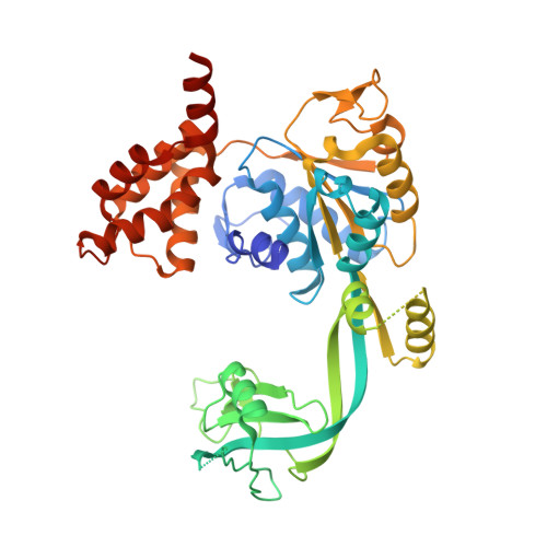
Crystal structure of the human AAA+ protein RuvBL1.

Matias, P.M.Gorynia, S.Donner, P.Carrondo, M.A.

(2006) J Biol Chem 281: 38918-38929

  • DOI: https://doi.org/10.1074/jbc.M605625200
  • Primary Citation of Related Structures:  
    2C9O

  • PubMed Abstract: 

    RuvBL1 is an evolutionarily highly conserved eukaryotic protein belonging to the AAA(+)-family of ATPases (ATPase associated with diverse cellular activities). It plays important roles in essential signaling pathways such as the c-Myc and Wnt pathways in chromatin remodeling, transcriptional and developmental regulation, and DNA repair and apoptosis. Herein we present the three-dimensional structure of the selenomethionine variant of human RuvBL1 refined using diffraction data to 2.2A of resolution. The crystal structure of the hexamer is formed of ADP-bound RuvBL1 monomers. The monomers contain three domains, of which the first and the third are involved in ATP binding and hydrolysis. Although it has been shown that ATPase activity of RuvBL1 is needed for several in vivo functions, we could only detect a marginal activity with the purified protein. Structural homology and DNA binding studies demonstrate that the second domain, which is unique among AAA(+) proteins and not present in the bacterial homolog RuvB, is a novel DNA/RNA-binding domain. We were able to demonstrate that RuvBL1 interacted with single-stranded DNA/RNA and double-stranded DNA. The structure of the RuvBL1.ADP complex, combined with our biochemical results, suggest that although RuvBL1 has all the structural characteristics of a molecular motor, even of an ATP-driven helicase, one or more as yet undetermined cofactors are needed for its enzymatic activity.


  • Organizational Affiliation

    Instituto de Tecnologia Química e Biológica, Universidade Nova de Lisboa, Apartado 127, 2781-901 Oeiras, Portugal.


Macromolecules
Find similar proteins by:  (by identity cutoff)  |  3D Structure
Entity ID: 1
MoleculeChains Sequence LengthOrganismDetailsImage
RUVB-LIKE 1
A, B, C
456Homo sapiensMutation(s): 0 
EC: 3.6.1
UniProt & NIH Common Fund Data Resources
Find proteins for Q9Y265 (Homo sapiens)
Explore Q9Y265 
Go to UniProtKB:  Q9Y265
PHAROS:  Q9Y265
GTEx:  ENSG00000175792 
Entity Groups  
Sequence Clusters30% Identity50% Identity70% Identity90% Identity95% Identity100% Identity
UniProt GroupQ9Y265
Sequence Annotations
Expand
  • Reference Sequence
Experimental Data & Validation

Experimental Data

  • Method: X-RAY DIFFRACTION
  • Resolution: 2.20 Å
  • R-Value Free: 0.257 
  • R-Value Work: 0.206 
  • R-Value Observed: 0.209 
  • Space Group: P 6
Unit Cell:
Length ( Å )Angle ( ˚ )
a = 207.08α = 90
b = 207.08β = 90
c = 60.71γ = 120
Software Package:
Software NamePurpose
SHARPmodel building
SCALAdata scaling
SHELXCDphasing
SHELXDphasing
SHELXEphasing
SHARPphasing
REFMACrefinement

Structure Validation

View Full Validation Report


Entry History 

Deposition Data

Revision History  (Full details and data files)

  • Version 1.0: 2006-10-23
    Type: Initial release
  • Version 1.1: 2011-05-08
    Changes: Version format compliance
  • Version 1.2: 2011-07-13
    Changes: Version format compliance
  • Version 1.3: 2018-03-28
    Changes: Database references, Source and taxonomy
  • Version 1.4: 2019-03-06
    Changes: Data collection, Experimental preparation
  • Version 1.5: 2019-05-08
    Changes: Data collection, Experimental preparation