2C8F

Structure of the ARTT motif E214N mutant C3bot1 Exoenzyme (NAD-bound state, crystal form III)


Experimental Data Snapshot

  • Method: X-RAY DIFFRACTION
  • Resolution: 2.50 Å
  • R-Value Free: 0.316 
  • R-Value Work: 0.232 
  • R-Value Observed: 0.237 

wwPDB Validation   3D Report Full Report


Ligand Structure Quality Assessment 


This is version 1.3 of the entry. See complete history


Literature

Structural Basis for the Nad-Hydrolysis Mechanism and the Artt-Loop Plasticity of C3 Exoenzymes.

Menetrey, J.Flatau, G.Boquet, P.Menez, A.Stura, E.A.

(2008) Protein Sci 17: 878

  • DOI: https://doi.org/10.1110/ps.073398508
  • Primary Citation of Related Structures:  
    2C89, 2C8A, 2C8B, 2C8C, 2C8D, 2C8E, 2C8F

  • PubMed Abstract: 

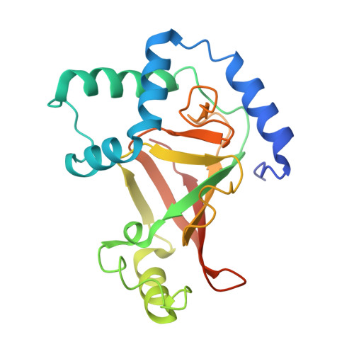
    C3-like exoenzymes are ADP-ribosyltransferases that specifically modify some Rho GTPase proteins, leading to their sequestration in the cytoplasm, and thus inhibiting their regulatory activity on the actin cytoskeleton. This modification process goes through three sequential steps involving NAD-hydrolysis, Rho recognition, and binding, leading to Rho ADP-ribosylation. Independently, three distinct residues within the ARTT loop of the C3 exoenzymes are critical for each of these steps. Supporting the critical role of the ARTT loop, we have shown previously that it adopts a distinct conformation upon NAD binding. Here, we present seven wild-type and ARTT loop-mutant structures of C3 exoenzyme of Clostridium botulinum free and bound to its true substrate, NAD, and to its NAD-hydrolysis product, nicotinamide. Altogether, these structures expand our understanding of the conformational diversity of the C3 exoenzyme, mainly within the ARTT loop.


  • Organizational Affiliation

    Institut Curie, Centre de Recherche, Paris F-75248, France. julie.Menetrey@curie.fr


Macromolecules
Find similar proteins by:  (by identity cutoff)  |  3D Structure
Entity ID: 1
MoleculeChains Sequence LengthOrganismDetailsImage
MONO-ADP-RIBOSYLTRANSFERASE C3A [auth E],
B [auth F],
C [auth G]
211Clostridium botulinumMutation(s): 1 
EC: 2.4.2
UniProt
Find proteins for Q7M0L1 (Clostridium botulinum)
Explore Q7M0L1 
Go to UniProtKB:  Q7M0L1
Entity Groups  
Sequence Clusters30% Identity50% Identity70% Identity90% Identity95% Identity100% Identity
UniProt GroupQ7M0L1
Sequence Annotations
Expand
  • Reference Sequence
Small Molecules
Ligands 1 Unique
IDChains Name / Formula / InChI Key2D Diagram3D Interactions
NAD
Query on NAD

Download Ideal Coordinates CCD File 
D [auth E]NICOTINAMIDE-ADENINE-DINUCLEOTIDE
C21 H27 N7 O14 P2
BAWFJGJZGIEFAR-NNYOXOHSSA-N
Experimental Data & Validation

Experimental Data

  • Method: X-RAY DIFFRACTION
  • Resolution: 2.50 Å
  • R-Value Free: 0.316 
  • R-Value Work: 0.232 
  • R-Value Observed: 0.237 
  • Space Group: P 1 21 1
Unit Cell:
Length ( Å )Angle ( ˚ )
a = 44.837α = 90
b = 70.381β = 97.28
c = 114.688γ = 90
Software Package:
Software NamePurpose
REFMACrefinement
DENZOdata reduction
SCALEPACKdata scaling
AMoREphasing

Structure Validation

View Full Validation Report


Ligand Structure Quality Assessment 


Entry History 

Deposition Data

Revision History  (Full details and data files)

  • Version 1.0: 2007-02-27
    Type: Initial release
  • Version 1.1: 2011-06-02
    Changes: Version format compliance
  • Version 1.2: 2011-07-13
    Changes: Version format compliance
  • Version 1.3: 2023-12-13
    Changes: Data collection, Database references, Derived calculations, Other, Refinement description