2C1U

CRYSTAL STRUCTURE OF THE DI-HAEM CYTOCHROME C PEROXIDASE FROM PARACOCCUS PANTOTROPHUS - OXIDISED FORM


Experimental Data Snapshot

  • Method: X-RAY DIFFRACTION
  • Resolution: 1.95 Å
  • R-Value Free: 0.220 
  • R-Value Work: 0.169 
  • R-Value Observed: 0.170 

wwPDB Validation   3D Report Full Report


This is version 1.3 of the entry. See complete history


Literature

Activation and Catalysis of the Di-Heme Cytochrome C Peroxidase from Paracoccus Pantotrophus

Echalier, A.Goodhew, C.F.Pettigrew, G.W.Fulop, V.

(2006) Structure 14: 107

  • DOI: https://doi.org/10.1016/j.str.2005.09.011
  • Primary Citation of Related Structures:  
    2C1U, 2C1V

  • PubMed Abstract: 

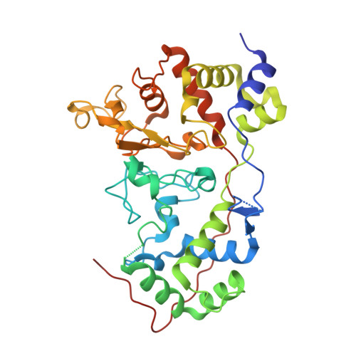
    Bacterial cytochrome c peroxidases contain an electron transferring (E) heme domain and a peroxidatic (P) heme domain. All but one of these enzymes are isolated in an inactive oxidized state and require reduction of the E heme by a small redox donor protein in order to activate the P heme. Here we present the structures of the inactive oxidized and active mixed valence enzyme from Paracoccus pantotrophus. Chain flexibility in the former, as expressed by the crystallographic temperature factors, is strikingly distributed in certain loop regions, and these coincide with the regions of conformational change that occur in forming the active mixed valence enzyme. On the basis of these changes, we postulate a series of events that occur to link the trigger of the electron entering the E heme from either pseudoazurin or cytochrome c(550) and the dissociation of a coordinating histidine at the P heme, which allows substrate access.


  • Organizational Affiliation

    Department of Biological Sciences, University of Warwick, Coventry CV4 7AL, UK.


Macromolecules
Find similar proteins by:  (by identity cutoff)  |  3D Structure
Entity ID: 1
MoleculeChains Sequence LengthOrganismDetailsImage
DI-HAEM CYTOCHROME C PEROXIDASE
A, B, C, D
338Paracoccus pantotrophusMutation(s): 0 
EC: 1.11.1.5
Entity Groups  
Sequence Clusters30% Identity50% Identity70% Identity90% Identity95% Identity100% Identity
Sequence Annotations
Expand
  • Reference Sequence
Small Molecules
Experimental Data & Validation

Experimental Data

  • Method: X-RAY DIFFRACTION
  • Resolution: 1.95 Å
  • R-Value Free: 0.220 
  • R-Value Work: 0.169 
  • R-Value Observed: 0.170 
  • Space Group: P 1 21 1
Unit Cell:
Length ( Å )Angle ( ˚ )
a = 78.8α = 90
b = 51.1β = 98.2
c = 167.9γ = 90
Software Package:
Software NamePurpose
REFMACrefinement
DENZOdata reduction
SCALEPACKdata scaling
AMoREphasing

Structure Validation

View Full Validation Report


Entry History 

Deposition Data

Revision History  (Full details and data files)

  • Version 1.0: 2006-01-13
    Type: Initial release
  • Version 1.1: 2011-05-08
    Changes: Version format compliance
  • Version 1.2: 2011-07-13
    Changes: Version format compliance
  • Version 1.3: 2023-12-13
    Changes: Data collection, Database references, Derived calculations, Other, Refinement description