2BX7

Crystal structure of L. lactis dihydroorotate dehydrogense A in complex with 3,5-dihydroxybenzoate


Experimental Data Snapshot

  • Method: X-RAY DIFFRACTION
  • Resolution: 2.04 Å
  • R-Value Free: 0.229 
  • R-Value Work: 0.184 
  • R-Value Observed: 0.187 

wwPDB Validation   3D Report Full Report


Ligand Structure Quality Assessment 


This is version 1.4 of the entry. See complete history


Literature

Interaction of Benzoate Pyrimidine Analogues with Class 1A Dihydroorotate Dehydrogenase from Lactococcus Lactis.

Wolfe, A.E.Thymark, M.Gattis, S.G.Fagan, R.L.Hu, Y.-C.Johansson, E.Arent, S.Larsen, S.Palfey, B.A.

(2007) Biochemistry 46: 5741

  • DOI: https://doi.org/10.1021/bi7001554
  • Primary Citation of Related Structures:  
    2BSL, 2BX7

  • PubMed Abstract: 

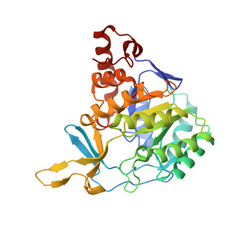
    Dihydroorotate dehydrogenases (DHODs) catalyze the oxidation of dihydroorotate to orotate in the only redox reaction in pyrimidine biosynthesis. The pyrimidine binding sites are very similar in all structurally characterized DHODs, suggesting that the prospects for identifying a class-specific inhibitor directed against this site are poor. Nonetheless, two compounds that bind specifically to the Class 1A DHOD from Lactococcus lactis, 3,4-dihydroxybenzoate (3,4-diOHB) and 3,5-dihydroxybenzoate (3,5-diOHB), have been identified [Palfey et al. (2001) J. Med. Chem. 44, 2861-2864]. The mechanism of inhibitor binding to the Class 1A DHOD from L. lactis has now been studied in detail and is reported here. Titrations showed that 3,4-diOHB binds more tightly at higher pH, whereas the opposite is true for 3,5-diOHB. Isothermal titration calorimetry and absorbance spectroscopy showed that 3,4-diOHB ionizes to the phenolate upon binding to the enzyme, but 3,5-diOHB does not. The charge-transfer band that forms in the 3,4-diOHB complex allowed the kinetics of binding to be observed in stopped-flow experiments. Binding was slow enough to observe from pH 6 to pH 8 and was (minimally) a two-step process consisting of the rapid formation of a complex that isomerized to the final charge-transfer complex. Orotate and 3,5-diOHB bind too quickly to follow directly, but their dissociation kinetics were studied by competition and described adequately with a single step. Crystal structures of both inhibitor complexes were determined, showing that 3,5-diOHB binds in the same orientation as orotate. In contrast, 3,4-diOHB binds in a twisted orientation, enabling one of its phenolic oxygens to form a very strong hydrogen bond to an asparagine, thus stabilizing the phenolate and causing charge-transfer interactions with the pi-system of the flavin, resulting in a green color.


  • Organizational Affiliation

    Department of Biological Chemistry, University of Michigan Medical School, 1150 West Medical Center Drive, Ann Arbor, Michigan 48109-0606, USA.


Macromolecules
Find similar proteins by:  (by identity cutoff)  |  3D Structure
Entity ID: 1
MoleculeChains Sequence LengthOrganismDetailsImage
DIHYDROOROTATE DEHYDROGENASE
A, B
311Lactococcus lactisMutation(s): 0 
EC: 1.3.3.1
UniProt
Find proteins for A2RJT9 (Lactococcus lactis subsp. cremoris (strain MG1363))
Explore A2RJT9 
Go to UniProtKB:  A2RJT9
Entity Groups  
Sequence Clusters30% Identity50% Identity70% Identity90% Identity95% Identity100% Identity
UniProt GroupA2RJT9
Sequence Annotations
Expand
  • Reference Sequence
Small Molecules
Ligands 5 Unique
IDChains Name / Formula / InChI Key2D Diagram3D Interactions
FMN
Query on FMN

Download Ideal Coordinates CCD File 
C [auth A],
G [auth B]
FLAVIN MONONUCLEOTIDE
C17 H21 N4 O9 P
FVTCRASFADXXNN-SCRDCRAPSA-N
34D
Query on 34D

Download Ideal Coordinates CCD File 
H [auth B]3,5-DIHYDROXYBENZOATE
C7 H6 O4
UYEMGAFJOZZIFP-UHFFFAOYSA-N
GOL
Query on GOL

Download Ideal Coordinates CCD File 
F [auth A]GLYCEROL
C3 H8 O3
PEDCQBHIVMGVHV-UHFFFAOYSA-N
ACT
Query on ACT

Download Ideal Coordinates CCD File 
D [auth A]ACETATE ION
C2 H3 O2
QTBSBXVTEAMEQO-UHFFFAOYSA-M
MG
Query on MG

Download Ideal Coordinates CCD File 
E [auth A],
I [auth B]
MAGNESIUM ION
Mg
JLVVSXFLKOJNIY-UHFFFAOYSA-N
Experimental Data & Validation

Experimental Data

  • Method: X-RAY DIFFRACTION
  • Resolution: 2.04 Å
  • R-Value Free: 0.229 
  • R-Value Work: 0.184 
  • R-Value Observed: 0.187 
  • Space Group: P 1 21 1
Unit Cell:
Length ( Å )Angle ( ˚ )
a = 53.71α = 90
b = 108.127β = 104.06
c = 66.216γ = 90
Software Package:
Software NamePurpose
REFMACrefinement
DENZOdata reduction
SCALEPACKdata scaling

Structure Validation

View Full Validation Report


Ligand Structure Quality Assessment 


Entry History 

Deposition Data

Revision History  (Full details and data files)

  • Version 1.0: 2006-08-29
    Type: Initial release
  • Version 1.1: 2011-05-08
    Changes: Version format compliance
  • Version 1.2: 2011-07-13
    Changes: Version format compliance
  • Version 1.3: 2019-05-08
    Changes: Data collection, Experimental preparation, Other
  • Version 1.4: 2023-12-13
    Changes: Data collection, Database references, Derived calculations, Other, Refinement description