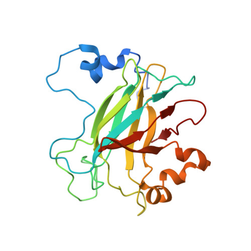
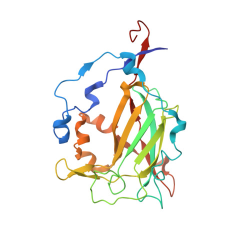
2BUQ

Crystal Structure Of Wild-Type Protocatechuate 3,4-Dioxygenase from Acinetobacter Sp. ADP1 in Complex with Catechol


Experimental Data Snapshot

  • Method: X-RAY DIFFRACTION
  • Resolution: 1.80 Å
  • R-Value Free: 0.194 
  • R-Value Work: 0.165 
  • R-Value Observed: 0.165 

wwPDB Validation   3D Report Full Report


This is version 1.5 of the entry. See complete history


Literature

Biophysical Analyses of Designed and Selected Mutants of Protocatechuate 3,4-Dioxygenase

Brown, C.K.Vetting, M.W.Earhart, C.A.Ohlendorf, D.H.

(2004) Annu Rev Microbiol 58: 555-585

  • DOI: https://doi.org/10.1146/annurev.micro.57.030502.090927
  • Primary Citation of Related Structures:  
    2BUM, 2BUQ, 2BUR, 2BUT, 2BUV

  • PubMed Abstract: 

    The catechol dioxygenases allow a wide variety of bacteria to use aromatic compounds as carbon sources by catalyzing the key ring-opening step. These enzymes use specifically either catechol or protocatechuate (2,3-dihydroxybenozate) as their substrates; they use a bare metal ion as the sole cofactor. To learn how this family of metalloenzymes functions, a structural analysis of designed and selected mutants of these enzymes has been undertaken. Here we review the results of this analysis on the nonheme ferric iron intradiol dioxygenase protocatechuate 3,4-dioxygenase.


  • Organizational Affiliation

    Center for Metals in Biocatalysis and Department of Biochemistry, Molecular Biology, and Biophysics , Minneapolis, Minnesota 55455, USA. brow0927@umn.edu


Macromolecules
Find similar proteins by:  (by identity cutoff)  |  3D Structure
Entity ID: 1
MoleculeChains Sequence LengthOrganismDetailsImage
PROTOCATECHUATE 3,4-DIOXYGENASE ALPHA CHAIN209Acinetobacter baylyi ADP1Mutation(s): 0 
EC: 1.13.11.3
UniProt
Find proteins for P20371 (Acinetobacter baylyi (strain ATCC 33305 / BD413 / ADP1))
Explore P20371 
Go to UniProtKB:  P20371
Entity Groups  
Sequence Clusters30% Identity50% Identity70% Identity90% Identity95% Identity100% Identity
UniProt GroupP20371
Sequence Annotations
Expand
  • Reference Sequence
Find similar proteins by:  (by identity cutoff)  |  3D Structure
Entity ID: 2
MoleculeChains Sequence LengthOrganismDetailsImage
PROTOCATECHUATE 3,4-DIOXYGENASE BETA CHAIN241Acinetobacter baylyi ADP1Mutation(s): 0 
EC: 1.13.11.3
UniProt
Find proteins for P20372 (Acinetobacter baylyi (strain ATCC 33305 / BD413 / ADP1))
Explore P20372 
Go to UniProtKB:  P20372
Entity Groups  
Sequence Clusters30% Identity50% Identity70% Identity90% Identity95% Identity100% Identity
UniProt GroupP20372
Sequence Annotations
Expand
  • Reference Sequence
Experimental Data & Validation

Experimental Data

  • Method: X-RAY DIFFRACTION
  • Resolution: 1.80 Å
  • R-Value Free: 0.194 
  • R-Value Work: 0.165 
  • R-Value Observed: 0.165 
  • Space Group: I 2 3
Unit Cell:
Length ( Å )Angle ( ˚ )
a = 144.7α = 90
b = 144.7β = 90
c = 144.7γ = 90
Software Package:
Software NamePurpose
CNSrefinement
CrystalCleardata reduction
CrystalCleardata scaling
CNSphasing

Structure Validation

View Full Validation Report


Entry History 

Deposition Data

Revision History  (Full details and data files)

  • Version 1.0: 2006-09-05
    Type: Initial release
  • Version 1.1: 2011-05-08
    Changes: Version format compliance
  • Version 1.2: 2011-07-13
    Changes: Version format compliance
  • Version 1.3: 2018-06-20
    Changes: Data collection, Database references
  • Version 1.4: 2019-05-22
    Changes: Data collection, Refinement description
  • Version 1.5: 2023-12-13
    Changes: Data collection, Database references, Derived calculations, Other, Refinement description