2BT1

Epstein Barr Virus dUTPase in complex with a,b-imino dUTP


Experimental Data Snapshot

  • Method: X-RAY DIFFRACTION
  • Resolution: 2.70 Å
  • R-Value Free: 0.245 
  • R-Value Work: 0.190 
  • R-Value Observed: 0.193 

wwPDB Validation   3D Report Full Report


Ligand Structure Quality Assessment 


This is version 1.3 of the entry. See complete history


Literature

The Monomeric Dutpase from Epstein-Barr Virus Mimics Trimeric Dutpases

Tarbouriech, N.Buisson, M.Seigneurin, J.-M.Cusack, S.Burmeister, W.P.

(2005) Structure 13: 1299

  • DOI: https://doi.org/10.1016/j.str.2005.06.009
  • Primary Citation of Related Structures:  
    2BSY, 2BT1

  • PubMed Abstract: 

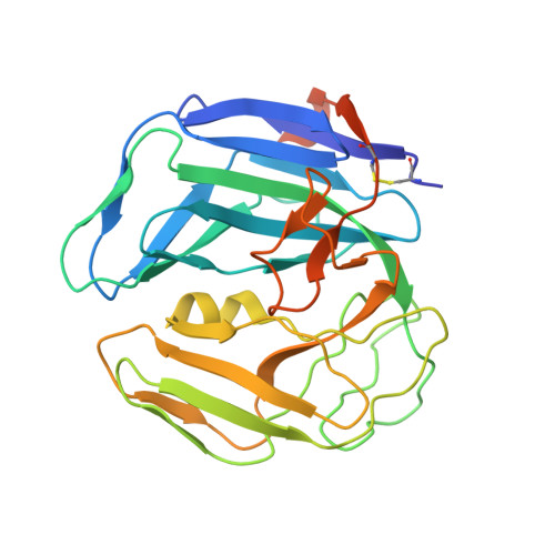
    Deoxyuridine 5'-triphosphate pyrophosphatases (dUTPases) are ubiquitous enzymes cleaving dUTP into dUMP and pyrophosphate. They occur as monomeric, dimeric, or trimeric molecules. The trimeric and monomeric enzymes both contain the same five characteristic sequence motifs but in a different order, whereas the dimeric enzymes are not homologous. Monomeric dUTPases only occur in herpesviruses, such as Epstein-Barr virus (EBV). Here, we describe the crystal structures of EBV dUTPase in complex with the product dUMP and a substrate analog alpha,beta-imino-dUTP. The molecule consists of three domains forming one active site that has a structure extremely similar to one of the three active sites of trimeric dUTPases. The three domains functionally correspond to the subunits of the trimeric form. Domains I and II have the dUTPase fold, but they differ considerably in the regions that are not involved in the formation of the unique active site, whereas domain III has only little secondary structure.


  • Organizational Affiliation

    EMBL-Grenoble Outstation, BP181, F-38042 Grenoble cedex 9, France. tarbour@embl-grenoble.fr


Macromolecules
Find similar proteins by:  (by identity cutoff)  |  3D Structure
Entity ID: 1
MoleculeChains Sequence LengthOrganismDetailsImage
DEOXYURIDINE 5'-TRIPHOSPHATE NUCLEOTIDOHYDROLASE278human gammaherpesvirus 4Mutation(s): 0 
EC: 3.6.1.23
UniProt
Find proteins for P03195 (Epstein-Barr virus (strain B95-8))
Explore P03195 
Go to UniProtKB:  P03195
Entity Groups  
Sequence Clusters30% Identity50% Identity70% Identity90% Identity95% Identity100% Identity
UniProt GroupP03195
Sequence Annotations
Expand
  • Reference Sequence
Small Molecules
Ligands 2 Unique
IDChains Name / Formula / InChI Key2D Diagram3D Interactions
DUP
Query on DUP

Download Ideal Coordinates CCD File 
B [auth A]2'-DEOXYURIDINE 5'-ALPHA,BETA-IMIDO-TRIPHOSPHATE
C9 H16 N3 O13 P3
XZLLMTSKYYYJLH-SHYZEUOFSA-N
MG
Query on MG

Download Ideal Coordinates CCD File 
C [auth A]MAGNESIUM ION
Mg
JLVVSXFLKOJNIY-UHFFFAOYSA-N
Experimental Data & Validation

Experimental Data

  • Method: X-RAY DIFFRACTION
  • Resolution: 2.70 Å
  • R-Value Free: 0.245 
  • R-Value Work: 0.190 
  • R-Value Observed: 0.193 
  • Space Group: P 62 2 2
Unit Cell:
Length ( Å )Angle ( ˚ )
a = 146.585α = 90
b = 146.585β = 90
c = 77.145γ = 120
Software Package:
Software NamePurpose
REFMACrefinement
MOSFLMdata reduction
SCALAdata scaling
MOLREPphasing

Structure Validation

View Full Validation Report


Ligand Structure Quality Assessment 


Entry History 

Deposition Data

Revision History  (Full details and data files)

  • Version 1.0: 2005-09-15
    Type: Initial release
  • Version 1.1: 2011-05-08
    Changes: Version format compliance
  • Version 1.2: 2011-07-13
    Changes: Version format compliance
  • Version 1.3: 2023-12-13
    Changes: Data collection, Database references, Derived calculations, Other, Refinement description