DNA Shuffling as a Tool for Protein Crystallization.
Keenan, R.J., Siehl, D.L., Gorton, R., Castle, L.A.(2005) Proc Natl Acad Sci U S A 102: 8887
- PubMed: 15951425
- DOI: https://doi.org/10.1073/pnas.0502497102
- Primary Citation of Related Structures:
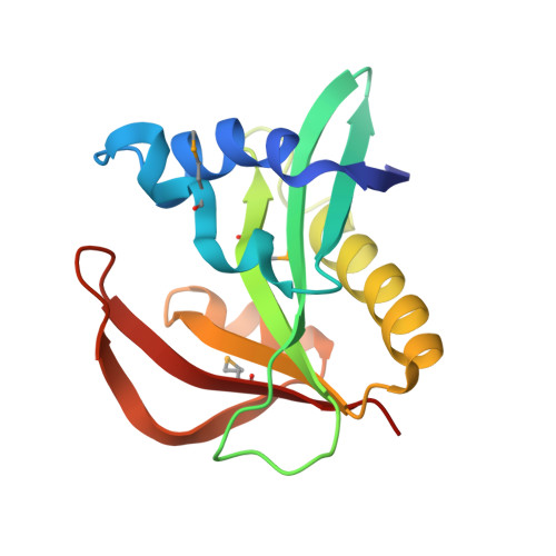
2BSW - PubMed Abstract:
The success of structural studies performed on an individual target in small scale or on many targets in the system-wide scale of structural genomics depends critically on three parameters: (i) obtaining an expression system capable of producing large quantities of the macromolecule(s) of interest, (ii) purifying this material in soluble form, and (iii) obtaining diffraction-quality crystals suitable for x-ray analysis. The attrition rate caused by these constraints is often quite high. Here, we present a strategy that addresses each of these three parameters simultaneously. Using DNA shuffling to introduce functional sequence variability into a protein of interest, we screened crude lysate supernatants for soluble variants that retain enzymatic activity. Crystallization trials performed on three WT and eight shuffled enzymes revealed two variants that crystallized readily. One of these was used to determine the high-resolution structure of the enzyme by x-ray analysis. The sequence diversity introduced through shuffling efficiently samples crystal packing space by modifying the surface properties of the enzyme. The approach demonstrated here does not require guidance as to the type of mutation necessary for improvements in expression, solubility, or crystallization. The method is scaleable and can be applied in situations where a single protein is being studied or in high-throughput structural genomics programs. Furthermore, it should be readily applied to structural studies of soluble proteins, membrane proteins, and macromolecular complexes.
Organizational Affiliation:
Pioneer Hi-Bred International, Inc., Verdia Campus, 700A Bay Road, Redwood City, CA 94063, USA. bkeenan@uchicago.edu