2APQ

Crystal Structure of an Active Site Mutant of Bovine Pancreatic Ribonuclease A (H119A-RNase A) with a 10-Glutamine expansion in the C-terminal hinge-loop.


Experimental Data Snapshot

  • Method: X-RAY DIFFRACTION
  • Resolution: 1.80 Å
  • R-Value Free: 0.215 
  • R-Value Work: 0.179 
  • R-Value Observed: 0.181 

wwPDB Validation   3D Report Full Report


This is version 1.4 of the entry. See complete history


Literature

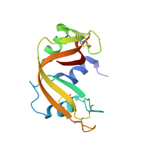
Amyloid-like fibrils of ribonuclease A with three-dimensional domain-swapped and native-like structure.

Sambashivan, S.Liu, Y.Sawaya, M.R.Gingery, M.Eisenberg, D.

(2005) Nature 437: 266-269

  • DOI: https://doi.org/10.1038/nature03916
  • Primary Citation of Related Structures:  
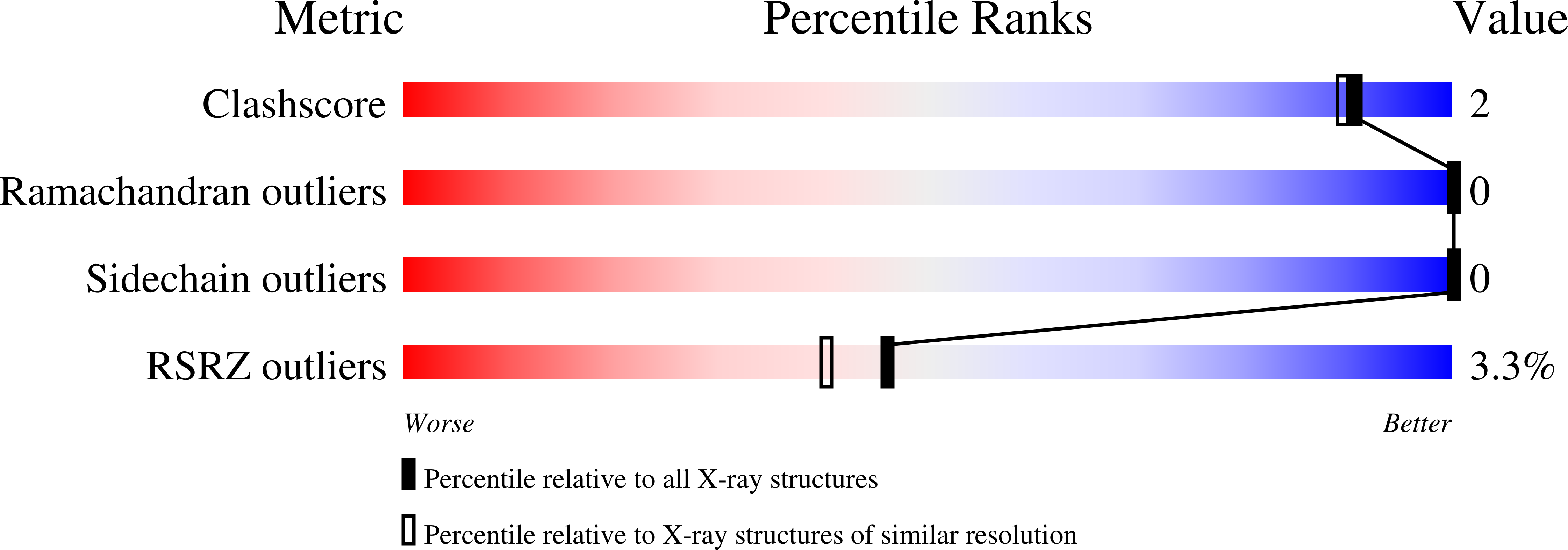
    2APQ

  • PubMed Abstract: 

    Amyloid or amyloid-like fibrils are elongated, insoluble protein aggregates, formed in vivo in association with neurodegenerative diseases or in vitro from soluble native proteins, respectively. The underlying structure of the fibrillar or 'cross-beta' state has presented long-standing, fundamental puzzles of protein structure. These include whether fibril-forming proteins have two structurally distinct stable states, native and fibrillar, and whether all or only part of the native protein refolds as it converts to the fibrillar state. Here we show that a designed amyloid-like fibril of the well-characterized enzyme RNase A contains native-like molecules capable of enzymatic activity. In addition, these functional molecular units are formed from a core RNase A domain and a swapped complementary domain. These findings are consistent with the zipper-spine model in which a cross-beta spine is decorated with three-dimensional domain-swapped functional units, retaining native-like structure.


  • Organizational Affiliation

    Howard Hughes Medical Institute, UCLA-DOE Institute for Genomics and Proteomics, Box 951570, UCLA, Los Angeles, California 90095-1570, USA.


Macromolecules
Find similar proteins by:  (by identity cutoff)  |  3D Structure
Entity ID: 1
MoleculeChains Sequence LengthOrganismDetailsImage
Ribonuclease139Bos taurusMutation(s): 1 
Gene Names: RNASE1RNS1
EC: 3.1.27.5
UniProt
Find proteins for P61823 (Bos taurus)
Explore P61823 
Go to UniProtKB:  P61823
Entity Groups  
Sequence Clusters30% Identity50% Identity70% Identity90% Identity95% Identity100% Identity
UniProt GroupP61823
Sequence Annotations
Expand
  • Reference Sequence
Experimental Data & Validation

Experimental Data

  • Method: X-RAY DIFFRACTION
  • Resolution: 1.80 Å
  • R-Value Free: 0.215 
  • R-Value Work: 0.179 
  • R-Value Observed: 0.181 
  • Space Group: P 32 2 1
Unit Cell:
Length ( Å )Angle ( ˚ )
a = 67.228α = 90
b = 67.228β = 90
c = 63.686γ = 120
Software Package:
Software NamePurpose
REFMACrefinement
CrystalCleardata reduction
SCALEPACKdata scaling
EPMRphasing

Structure Validation

View Full Validation Report


Entry History 

Deposition Data

Revision History  (Full details and data files)

  • Version 1.0: 2005-09-13
    Type: Initial release
  • Version 1.1: 2008-04-30
    Changes: Version format compliance
  • Version 1.2: 2011-07-13
    Changes: Version format compliance
  • Version 1.3: 2021-10-20
    Changes: Database references, Derived calculations
  • Version 1.4: 2023-08-23
    Changes: Data collection, Refinement description