Calorimetric and crystallographic analysis of the oligomeric structure of Escherichia coli GMP kinase
Hible, G., Renault, L., Schaeffer, F., Christova, P., Radulescu, A.Z., Evrin, C., Gilles, A.M., Cherfils, J.(2005) J Mol Biol 352: 1044-1059
- PubMed: 16140325
- DOI: https://doi.org/10.1016/j.jmb.2005.07.042
- Primary Citation of Related Structures:
2AN9, 2ANB, 2ANC - PubMed Abstract:
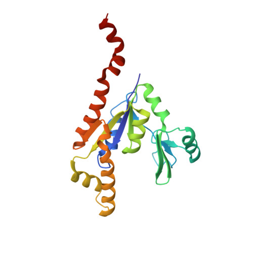
Guanosine monophosphate kinases (GMPKs), which catalyze the phosphorylation of GMP and dGMP to their diphosphate form, have been characterized as monomeric enzymes in eukaryotes and prokaryotes. Here, we report that GMPK from Escherichia coli (ecGMPK) assembles in solution and in the crystal as several different oligomers. Thermodynamic analysis of ecGMPK using differential scanning calorimetry shows that the enzyme is in equilibrium between a dimer and higher order oligomers, whose relative amounts depend on protein concentration, ionic strength, and the presence of ATP. Crystallographic structures of ecGMPK in the apo, GMP and GDP-bound forms were solved at 3.2A, 2.9A and 2.4A resolution, respectively. ecGMPK forms a hexamer with D3 symmetry in all crystal forms, in which the two nucleotide-binding domains are able to undergo closure comparable to that of monomeric GMPKs. The 2-fold and 3-fold interfaces involve a 20-residue C-terminal extension and a sequence signature, respectively, that are missing from monomeric eukaryotic GMPKs, explaining why ecGMPK forms oligomers. These signatures are found in GMPKs from proteobacteria, some of which are human pathogens. GMPKs from these bacteria are thus likely to form the same quaternary structures. The shift of the thermodynamic equilibrium towards the dimer at low ecGMPK concentration together with the observation that inter-subunit interactions partially occlude the ATP-binding site in the hexameric structure suggest that the dimer may be the active species at physiological enzyme concentration.
Organizational Affiliation:
Laboratoire d'Enzymologie et Biochimie Structurales, CNRS, Gif sur Yvette 91198, France.