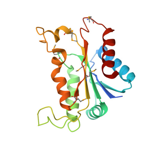
2AMJ

Crystal Structure of Modulator of Drug Activity B from Escherichia coli O157:H7


Experimental Data Snapshot

  • Method: X-RAY DIFFRACTION
  • Resolution: 1.80 Å
  • R-Value Free: 0.263 
  • R-Value Work: 0.233 
  • R-Value Observed: 0.233 

wwPDB Validation   3D Report Full Report


This is version 1.3 of the entry. See complete history


Literature

Modulator of drug activity B from Escherichia coli: crystal structure of a prokaryotic homologue of DT-diaphorase.

Adams, M.A.Jia, Z.

(2006) J Mol Biol 359: 455-465

  • DOI: https://doi.org/10.1016/j.jmb.2006.03.053
  • Primary Citation of Related Structures:  
    2AMJ, 2B3D

  • PubMed Abstract: 

    Modulator of drug activity B (MdaB) is a putative member of the DT-diaphorase family of NAD(P)H:oxidoreductases that afford cellular protection against quinonoid compounds. While there have been extensive investigations of mammalian homologues, putative prokaryotic members of this enzyme family have received little attention. The three-dimensional crystal structure of apo-MdaB reported herein exhibits significant structural similarity to a number of flavoproteins, including the mammalian DT-diaphorases. We have shown by mass spectrometry that the endogenously associated cofactor is flavin adenine dinucleotide and we present here the structure of MdaB in complex with this compound. Growth of Escherichia coli carrying null mutations in the genes encoding MdaB or quinol monooxygenase, the gene for which shares the mdaB promoter, were not affected by the presence of menadione. However, over-expression of recombinant quinol monooxygenase conferred a state of resistance against both tetracycline and adriamycin. This work suggests that the redox cycle formed by these proteins protects E. coli from the toxic effects of polyketide compounds rather than the oxidative stress of menadione alone.


  • Organizational Affiliation

    Department of Biochemistry, Queen's University, Kingston, Ont., Canada K7L 3N6.


Macromolecules
Find similar proteins by:  (by identity cutoff)  |  3D Structure
Entity ID: 1
MoleculeChains Sequence LengthOrganismDetailsImage
Modulator of drug activity B
A, B, C, D
204Escherichia coli O157:H7Mutation(s): 6 
Gene Names: mdaBmda66
UniProt
Find proteins for P0AEY7 (Escherichia coli O157:H7)
Explore P0AEY7 
Go to UniProtKB:  P0AEY7
Entity Groups  
Sequence Clusters30% Identity50% Identity70% Identity90% Identity95% Identity100% Identity
UniProt GroupP0AEY7
Sequence Annotations
Expand
  • Reference Sequence
Small Molecules
Modified Residues  1 Unique
IDChains TypeFormula2D DiagramParent
MSE
Query on MSE
A, B, C, D
L-PEPTIDE LINKINGC5 H11 N O2 SeMET
Experimental Data & Validation

Experimental Data

  • Method: X-RAY DIFFRACTION
  • Resolution: 1.80 Å
  • R-Value Free: 0.263 
  • R-Value Work: 0.233 
  • R-Value Observed: 0.233 
  • Space Group: P 1 21 1
Unit Cell:
Length ( Å )Angle ( ˚ )
a = 53.056α = 90
b = 86.51β = 93.85
c = 82.667γ = 90
Software Package:
Software NamePurpose
HKL-2000data collection
SCALEPACKdata scaling
SHELXmodel building
SOLVEphasing
CNSrefinement
HKL-2000data reduction
SHELXphasing

Structure Validation

View Full Validation Report


Entry History 

Deposition Data

Revision History  (Full details and data files)

  • Version 1.0: 2006-07-18
    Type: Initial release
  • Version 1.1: 2008-04-30
    Changes: Version format compliance
  • Version 1.2: 2011-07-13
    Changes: Source and taxonomy, Version format compliance
  • Version 1.3: 2017-10-11
    Changes: Refinement description