Characterization of human DHRS6, an orphan short chain dehydrogenase/reductase enzyme: a novel, cytosolic type 2 R-beta-hydroxybutyrate dehydrogenase
Guo, K., Lukacik, P., Papagrigoriou, E., Meier, M., Lee, W.H., Adamski, J., Oppermann, U.(2006) J Biol Chem 281: 10291-10297
- PubMed: 16380372
- DOI: https://doi.org/10.1074/jbc.M511346200
- Primary Citation of Related Structures:
2AG5 - PubMed Abstract:
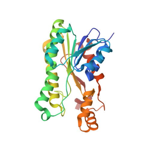
Human DHRS6 is a previously uncharacterized member of the short chain dehydrogenases/reductase family and displays significant homologies to bacterial hydroxybutyrate dehydrogenases. Substrate screening reveals sole NAD(+)-dependent conversion of (R)-hydroxybutyrate to acetoacetate with K(m) values of about 10 mm, consistent with plasma levels of circulating ketone bodies in situations of starvation or ketoacidosis. The structure of human DHRS6 was determined at a resolution of 1.8 A in complex with NAD(H) and reveals a tetrameric organization with a short chain dehydrogenases/reductase-typical folding pattern. A highly conserved triad of Arg residues ("triple R" motif consisting of Arg(144), Arg(188), and Arg(205)) was found to bind a sulfate molecule at the active site. Docking analysis of R-beta-hydroxybutyrate into the active site reveals an experimentally consistent model of substrate carboxylate binding and catalytically competent orientation. GFP reporter gene analysis reveals a cytosolic localization upon transfection into mammalian cells. These data establish DHRS6 as a novel, cytosolic type 2 (R)-hydroxybutyrate dehydrogenase, distinct from its well characterized mitochondrial type 1 counterpart. The properties determined for DHRS6 suggest a possible physiological role in cytosolic ketone body utilization, either as a secondary system for energy supply in starvation or to generate precursors for lipid and sterol synthesis.
Organizational Affiliation:
Structural Genomics Consortium, University of Oxford, Oxford OX3 7LD, United Kingdom.