2ZDI

Crystal structure of Prefoldin from Pyrococcus horikoshii OT3


Experimental Data Snapshot

  • Method: X-RAY DIFFRACTION
  • Resolution: 3.00 Å
  • R-Value Free: 0.277 
  • R-Value Work: 0.240 

wwPDB Validation   3D Report Full Report


This is version 1.2 of the entry. See complete history


Literature

Structure and molecular dynamics simulation of archaeal prefoldin: the molecular mechanism for binding and recognition of nonnative substrate proteins

Ohtaki, A.Kida, H.Miyata, Y.Ide, N.Yonezawa, A.Arakawa, T.Iizuka, R.Noguchi, K.Kita, A.Odaka, M.Miki, K.Yohda, M.

(2008) J Mol Biol 376: 1130-1141

  • DOI: https://doi.org/10.1016/j.jmb.2007.12.010
  • Primary Citation of Related Structures:  
    2ZDI

  • PubMed Abstract: 

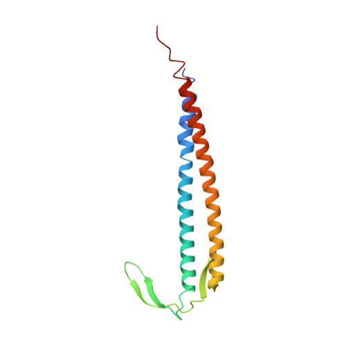
    Prefoldin (PFD) is a heterohexameric molecular chaperone complex in the eukaryotic cytosol and archaea with a jellyfish-like structure containing six long coiled-coil tentacles. PFDs capture protein folding intermediates or unfolded polypeptides and transfer them to group II chaperonins for facilitated folding. Although detailed studies on the mechanisms for interaction with unfolded proteins or cooperation with chaperonins of archaeal PFD have been performed, it is still unclear how PFD captures the unfolded protein. In this study, we determined the X-ray structure of Pyrococcus horikoshii OT3 PFD (PhPFD) at 3.0 A resolution and examined the molecular mechanism for binding and recognition of nonnative substrate proteins by molecular dynamics (MD) simulation and mutation analyses. PhPFD has a jellyfish-like structure with six long coiled-coil tentacles and a large central cavity. Each subunit has a hydrophobic groove at the distal region where an unfolded substrate protein is bound. During MD simulation at 330 K, each coiled coil was highly flexible, enabling it to widen its central cavity and capture various nonnative proteins. Docking MD simulation of PhPFD with unfolded insulin showed that the beta subunit is essentially involved in substrate binding and that the alpha subunit modulates the shape and width of the central cavity. Analyses of mutant PhPFDs with amino acid replacement of the hydrophobic residues of the beta subunit in the hydrophobic groove have shown that beta Ile107 has a critical role in forming the hydrophobic groove.


  • Organizational Affiliation

    Department of Biotechnology and Life Science, Tokyo University of Agriculture and Technology, 2-24-16 Naka-cho, Koganei, Tokyo 184-8588, Japan.


Macromolecules
Find similar proteins by:  (by identity cutoff)  |  3D Structure
Entity ID: 1
MoleculeChains Sequence LengthOrganismDetailsImage
Prefoldin subunit beta
A, B
117Pyrococcus horikoshiiMutation(s): 0 
Gene Names: pfdB
UniProt
Find proteins for O58268 (Pyrococcus horikoshii (strain ATCC 700860 / DSM 12428 / JCM 9974 / NBRC 100139 / OT-3))
Explore O58268 
Go to UniProtKB:  O58268
Entity Groups  
Sequence Clusters30% Identity50% Identity70% Identity90% Identity95% Identity100% Identity
UniProt GroupO58268
Sequence Annotations
Expand
  • Reference Sequence
Find similar proteins by:  (by identity cutoff)  |  3D Structure
Entity ID: 2
MoleculeChains Sequence LengthOrganismDetailsImage
Prefoldin subunit alpha151Pyrococcus horikoshiiMutation(s): 0 
Gene Names: pfdA
UniProt
Find proteins for O58263 (Pyrococcus horikoshii (strain ATCC 700860 / DSM 12428 / JCM 9974 / NBRC 100139 / OT-3))
Explore O58263 
Go to UniProtKB:  O58263
Entity Groups  
Sequence Clusters30% Identity50% Identity70% Identity90% Identity95% Identity100% Identity
UniProt GroupO58263
Sequence Annotations
Expand
  • Reference Sequence
Experimental Data & Validation

Experimental Data

  • Method: X-RAY DIFFRACTION
  • Resolution: 3.00 Å
  • R-Value Free: 0.277 
  • R-Value Work: 0.240 
  • Space Group: P 21 21 2
Unit Cell:
Length ( Å )Angle ( ˚ )
a = 65.173α = 90
b = 98.75β = 90
c = 78.615γ = 90
Software Package:
Software NamePurpose
HKL-2000data collection
MOLREPphasing
PHENIXrefinement
HKL-2000data reduction
HKL-2000data scaling

Structure Validation

View Full Validation Report


Entry History 

Deposition Data

Revision History  (Full details and data files)

  • Version 1.0: 2008-02-05
    Type: Initial release
  • Version 1.1: 2011-07-13
    Changes: Version format compliance
  • Version 1.2: 2023-11-01
    Changes: Data collection, Database references, Derived calculations, Refinement description