2Z32

Crystal structure of Keap1 complexed with Prothymosin alpha


Experimental Data Snapshot

  • Method: X-RAY DIFFRACTION
  • Resolution: 2.00 Å
  • R-Value Free: 0.223 
  • R-Value Work: 0.166 
  • R-Value Observed: 0.169 

wwPDB Validation   3D Report Full Report


This is version 1.2 of the entry. See complete history


Literature

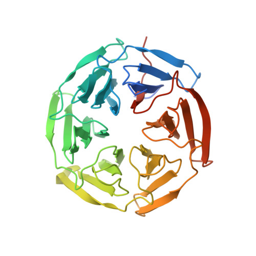
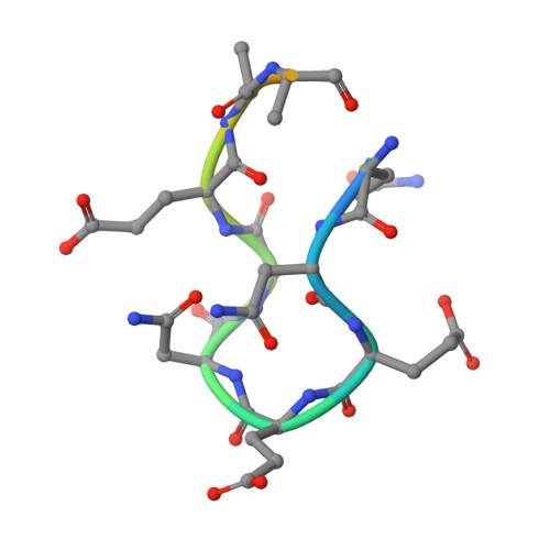
Structural analysis of the complex of Keap1 with a prothymosin alpha peptide

Padmanabhan, B.Nakamura, Y.Yokoyama, S.

(2008) Acta Crystallogr Sect F Struct Biol Cryst Commun 64: 233-238

  • DOI: https://doi.org/10.1107/S1744309108004995
  • Primary Citation of Related Structures:  
    2Z32

  • PubMed Abstract: 

    The Nrf2 transcription factor, which plays important roles in oxidative and xenobiotic stress, is negatively regulated by the cytoplasmic repressor Keap1. The beta-propeller/Kelch domain of Keap1, which is formed by the double-glycine repeat and C-terminal region domains (Keap1-DC), interacts directly with the Neh2 domain of Nrf2. The nuclear oncoprotein prothymosin alpha (ProTalpha) also interacts directly with Keap1 and may play a role in the dissociation of the Keap1-Nrf2 complex. The structure of Keap1-DC complexed with a ProTalpha peptide (amino acids 39-54) has been determined at 1.9 A resolution. The Keap1-bound ProTalpha peptide possesses a hairpin conformation and binds to the Keap1 protein at the bottom region of the beta-propeller domain. Complex formation occurs as a consequence of their complementary electrostatic interactions. A comparison of the present structure with recently reported Keap1-DC complex structures revealed that the DLG and ETGE motifs of the Neh2 domain of Nrf2 and the ProTalpha peptide bind to Keap1 in a similar manner but with different binding potencies.


  • Organizational Affiliation

    Genomic Sciences Center, Yokohama Institute, RIKEN, 1-7-22 Suehiro-cho, Tsurumi, Yokohama 230-0045, Japan. paddy@gsc.riken.jp


Macromolecules
Find similar proteins by:  (by identity cutoff)  |  3D Structure
Entity ID: 1
MoleculeChains Sequence LengthOrganismDetailsImage
Kelch-like ECH-associated protein 1318Mus musculusMutation(s): 0 
Gene Names: Keap1
UniProt
Find proteins for Q9Z2X8 (Mus musculus)
Explore Q9Z2X8 
Go to UniProtKB:  Q9Z2X8
Entity Groups  
Sequence Clusters30% Identity50% Identity70% Identity90% Identity95% Identity100% Identity
UniProt GroupQ9Z2X8
Sequence Annotations
Expand
  • Reference Sequence

Find similar proteins by:  Sequence   |   3D Structure  

Entity ID: 2
MoleculeChains Sequence LengthOrganismDetailsImage
Prothymosin alpha16N/AMutation(s): 0 
UniProt & NIH Common Fund Data Resources
Find proteins for P26350 (Mus musculus)
Explore P26350 
Go to UniProtKB:  P26350
IMPC:  MGI:97803
Entity Groups  
Sequence Clusters30% Identity50% Identity70% Identity90% Identity95% Identity100% Identity
UniProt GroupP26350
Sequence Annotations
Expand
  • Reference Sequence
Experimental Data & Validation

Experimental Data

  • Method: X-RAY DIFFRACTION
  • Resolution: 2.00 Å
  • R-Value Free: 0.223 
  • R-Value Work: 0.166 
  • R-Value Observed: 0.169 
  • Space Group: P 61
Unit Cell:
Length ( Å )Angle ( ˚ )
a = 103.515α = 90
b = 103.515β = 90
c = 56.16γ = 120
Software Package:
Software NamePurpose
REFMACrefinement
ADSCdata collection
HKL-2000data reduction
SCALEPACKdata scaling
MOLREPphasing

Structure Validation

View Full Validation Report


Entry History 

Deposition Data

Revision History  (Full details and data files)

  • Version 1.0: 2008-03-11
    Type: Initial release
  • Version 1.1: 2011-07-13
    Changes: Version format compliance
  • Version 1.2: 2023-11-01
    Changes: Data collection, Database references, Derived calculations, Refinement description