A Glutathione Transferase from Agrobacterium Tumefaciens Reveals a Novel Class of Bacterial Gst Superfamily.
Skopelitou, K., Dhavala, P., Papageorgiou, A.C., Labrou, N.E.(2012) PLoS One 7: 34263
- PubMed: 22496785
- DOI: https://doi.org/10.1371/journal.pone.0034263
- Primary Citation of Related Structures:
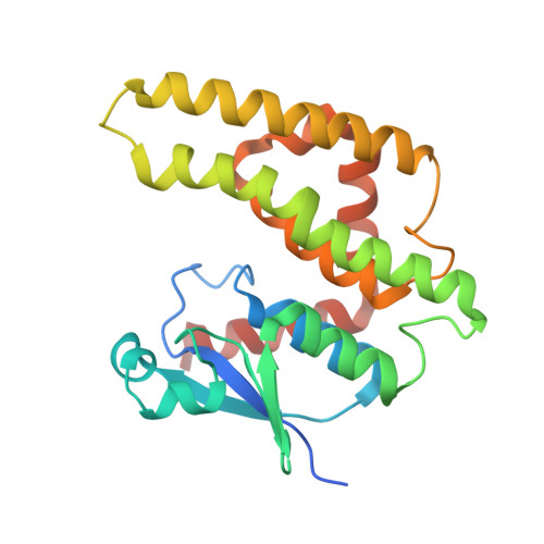
2YCD - PubMed Abstract:
In the present work, we report a novel class of glutathione transferases (GSTs) originated from the pathogenic soil bacterium Agrobacterium tumefaciens C58, with structural and catalytic properties not observed previously in prokaryotic and eukaryotic GST isoenzymes. A GST-like sequence from A. tumefaciens C58 (Atu3701) with low similarity to other characterized GST family of enzymes was identified. Phylogenetic analysis showed that it belongs to a distinct GST class not previously described and restricted only in soil bacteria, called the Eta class (H). This enzyme (designated as AtuGSTH1-1) was cloned and expressed in E. coli and its structural and catalytic properties were investigated. Functional analysis showed that AtuGSTH1-1 exhibits significant transferase activity against the common substrates aryl halides, as well as very high peroxidase activity towards organic hydroperoxides. The crystal structure of AtuGSTH1-1 was determined at 1.4 Å resolution in complex with S-(p-nitrobenzyl)-glutathione (Nb-GSH). Although AtuGSTH1-1 adopts the canonical GST fold, sequence and structural characteristics distinct from previously characterized GSTs were identified. The absence of the classic catalytic essential residues (Tyr, Ser, Cys) distinguishes AtuGSTH1-1 from all other cytosolic GSTs of known structure and function. Site-directed mutagenesis showed that instead of the classic catalytic residues, an Arg residue (Arg34), an electron-sharing network, and a bridge of a network of water molecules may form the basis of the catalytic mechanism. Comparative sequence analysis, structural information, and site-directed mutagenesis in combination with kinetic analysis showed that Phe22, Ser25, and Arg187 are additional important residues for the enzyme's catalytic efficiency and specificity.
Organizational Affiliation:
Laboratory of Enzyme Technology, Department of Agricultural Biotechnology, Agricultural University of Athens, Athens, Greece.