2Y48

Crystal structure of LSD1-CoREST in complex with a N-terminal SNAIL peptide


Experimental Data Snapshot

  • Method: X-RAY DIFFRACTION
  • Resolution: 3.00 Å
  • R-Value Free: 0.246 
  • R-Value Work: 0.212 
  • R-Value Observed: 0.213 

wwPDB Validation   3D Report Full Report


Ligand Structure Quality Assessment 


This is version 1.3 of the entry. See complete history


Literature

Molecular Mimicry and Ligand Recognition in Binding and Catalysis by the Histone Demethylase Lsd1-Corest Complex.

Baron, R.Binda, C.Tortorici, M.Mccammon, J.A.Mattevi, A.

(2011) Structure 19: 212

  • DOI: https://doi.org/10.1016/j.str.2011.01.001
  • Primary Citation of Related Structures:  
    2Y48

  • PubMed Abstract: 

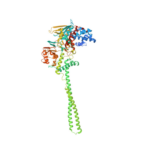
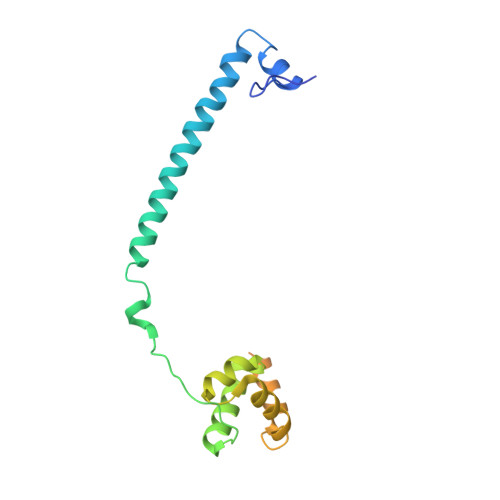
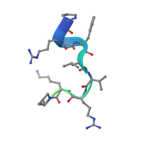
    Histone demethylases LSD1 and LSD2 (KDM1A/B) catalyze the oxidative demethylation of Lys4 of histone H3. We used molecular dynamics simulations to probe the diffusion of the oxygen substrate. Oxygen can reach the catalytic center independently from the presence of a bound histone peptide, implying that LSD1 can complete subsequent demethylation cycles without detaching from the nucleosomal particle. The simulations highlight the role of a strictly conserved active-site Lys residue providing general insight into the enzymatic mechanism of oxygen-reacting flavoenzymes. The crystal structure of LSD1-CoREST bound to a peptide of the transcription factor SNAIL1 unravels a fascinating example of molecular mimicry. The SNAIL1 N-terminal residues bind to the enzyme active-site cleft, effectively mimicking the H3 tail. This finding predicts that other members of the SNAIL/Scratch transcription factor family might associate to LSD1/2. The combination of selective histone-modifying activity with the distinct recognition mechanisms underlies the biological complexity of LSD1/2.


  • Organizational Affiliation

    Department of Chemistry and Biochemistry, Center for Theoretical Biological Physics, and Department of Pharmacology, Howard Hughes Medical Institute, University of California at San Diego, La Jolla, CA 92093-0365, USA. rbaron@mccammon.ucsd.edu


Macromolecules
Find similar proteins by:  (by identity cutoff)  |  3D Structure
Entity ID: 1
MoleculeChains Sequence LengthOrganismDetailsImage
LYSINE-SPECIFIC DEMETHYLASE 1A730Homo sapiensMutation(s): 0 
EC: 1
UniProt & NIH Common Fund Data Resources
Find proteins for O60341 (Homo sapiens)
Explore O60341 
Go to UniProtKB:  O60341
PHAROS:  O60341
GTEx:  ENSG00000004487 
Entity Groups  
Sequence Clusters30% Identity50% Identity70% Identity90% Identity95% Identity100% Identity
UniProt GroupO60341
Sequence Annotations
Expand
  • Reference Sequence
Find similar proteins by:  (by identity cutoff)  |  3D Structure
Entity ID: 2
MoleculeChains Sequence LengthOrganismDetailsImage
REST COREPRESSOR 1178Homo sapiensMutation(s): 0 
UniProt & NIH Common Fund Data Resources
Find proteins for Q9UKL0 (Homo sapiens)
Explore Q9UKL0 
Go to UniProtKB:  Q9UKL0
PHAROS:  Q9UKL0
GTEx:  ENSG00000089902 
Entity Groups  
Sequence Clusters30% Identity50% Identity70% Identity90% Identity95% Identity100% Identity
UniProt GroupQ9UKL0
Sequence Annotations
Expand
  • Reference Sequence

Find similar proteins by:  Sequence   |   3D Structure  

Entity ID: 3
MoleculeChains Sequence LengthOrganismDetailsImage
ZINC FINGER PROTEIN SNAI120Homo sapiensMutation(s): 0 
UniProt & NIH Common Fund Data Resources
Find proteins for O95863 (Homo sapiens)
Explore O95863 
Go to UniProtKB:  O95863
PHAROS:  O95863
GTEx:  ENSG00000124216 
Entity Groups  
Sequence Clusters30% Identity50% Identity70% Identity90% Identity95% Identity100% Identity
UniProt GroupO95863
Sequence Annotations
Expand
  • Reference Sequence
Small Molecules
Ligands 1 Unique
IDChains Name / Formula / InChI Key2D Diagram3D Interactions
FAD
Query on FAD

Download Ideal Coordinates CCD File 
D [auth A]FLAVIN-ADENINE DINUCLEOTIDE
C27 H33 N9 O15 P2
VWWQXMAJTJZDQX-UYBVJOGSSA-N
Experimental Data & Validation

Experimental Data

  • Method: X-RAY DIFFRACTION
  • Resolution: 3.00 Å
  • R-Value Free: 0.246 
  • R-Value Work: 0.212 
  • R-Value Observed: 0.213 
  • Space Group: I 2 2 2
Unit Cell:
Length ( Å )Angle ( ˚ )
a = 119.17α = 90
b = 181.46β = 90
c = 234.44γ = 90
Software Package:
Software NamePurpose
REFMACrefinement
MOSFLMdata reduction
SCALAdata scaling
AMoREphasing

Structure Validation

View Full Validation Report


Ligand Structure Quality Assessment 


Entry History 

Deposition Data

Revision History  (Full details and data files)

  • Version 1.0: 2011-02-16
    Type: Initial release
  • Version 1.1: 2011-05-08
    Changes: Version format compliance
  • Version 1.2: 2011-07-13
    Changes: Version format compliance
  • Version 1.3: 2023-12-20
    Changes: Data collection, Database references, Derived calculations, Other, Refinement description