2XX9

Crystal structure of 1-((2-fluoro-4-(3-(trifluoromethyl)-4,5,6,7- tetrahydro-1H-indazol-1-yl)phenyl)methyl)-2-pyrrolidinone in complex with the ligand binding domain of the Rat GluA2 receptor and glutamate at 2.2A resolution.


Experimental Data Snapshot

  • Method: X-RAY DIFFRACTION
  • Resolution: 1.97 Å
  • R-Value Free: 0.224 
  • R-Value Work: 0.185 
  • R-Value Observed: 0.187 

wwPDB Validation   3D Report Full Report


Ligand Structure Quality Assessment 


This is version 1.1 of the entry. See complete history


Literature

Integration of Lead Optimization with Crystallography for a Membrane-Bound Ion Channel Target: Discovery of a New Class of Ampa Receptor Positive Allosteric Modulators.

Ward, S.E.Harries, M.Aldegheri, L.Austin, N.E.Ballantine, S.Ballini, E.Bradley, D.M.Bax, B.D.Clarke, B.P.Harris, A.J.Harrison, S.A.Melarange, R.A.Mookherjee, C.Mosley, J.Dal Negro, G.Oliosi, B.Smith, K.J.Thewlis, K.M.Woollard, P.M.Yusaf, S.P.

(2011) J Med Chem 54: 78

  • DOI: https://doi.org/10.1021/jm100679e
  • Primary Citation of Related Structures:  
    2XX7, 2XX8, 2XX9, 2XXH, 2XXI

  • PubMed Abstract: 

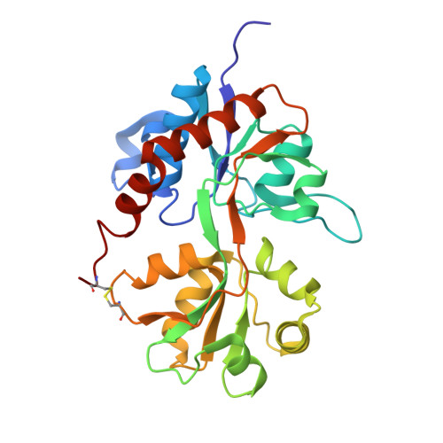
    A novel series of AMPAR positive modulators is described that were identified by high throughput screening. The molecules of the series have been optimized from a high quality starting point hit to afford excellent developability, tolerability, and efficacy profiles, leading to identification of a clinical candidate. Unusually for an ion channel target, this optimization was integrated with regular generation of ligand-bound crystal structures and uncovered a novel chemotype with a unique and highly conserved mode of interaction via a trifluoromethyl group.


  • Organizational Affiliation

    School of Life Sciences, University of Sussex, Brighton, United Kingdom. simon.ward@sussex.ac.uk


Macromolecules
Find similar proteins by:  (by identity cutoff)  |  3D Structure
Entity ID: 1
MoleculeChains Sequence LengthOrganismDetailsImage
GLUTAMATE RECEPTOR 2
A, B, C
263Rattus norvegicusMutation(s): 1 
UniProt
Find proteins for P19491 (Rattus norvegicus)
Explore P19491 
Go to UniProtKB:  P19491
Entity Groups  
Sequence Clusters30% Identity50% Identity70% Identity90% Identity95% Identity100% Identity
UniProt GroupP19491
Sequence Annotations
Expand
  • Reference Sequence
Small Molecules
Ligands 4 Unique
IDChains Name / Formula / InChI Key2D Diagram3D Interactions
1NF
Query on 1NF

Download Ideal Coordinates CCD File 
E [auth A],
I [auth B]
N,N-DIMETHYL-4-[3-(TRIFLUOROMETHYL)-4,5,6,7-TETRAHYDRO-1H-INDAZOL-1-YL]BENZAMIDE
C19 H19 F4 N3 O
FMFRNCRYWTXTOL-UHFFFAOYSA-N
GLU
Query on GLU

Download Ideal Coordinates CCD File 
D [auth A],
H [auth B],
L [auth C]
GLUTAMIC ACID
C5 H9 N O4
WHUUTDBJXJRKMK-VKHMYHEASA-N
SO4
Query on SO4

Download Ideal Coordinates CCD File 
F [auth A]SULFATE ION
O4 S
QAOWNCQODCNURD-UHFFFAOYSA-L
ZN
Query on ZN

Download Ideal Coordinates CCD File 
G [auth A],
J [auth B],
K [auth B],
M [auth C],
N [auth C]
ZINC ION
Zn
PTFCDOFLOPIGGS-UHFFFAOYSA-N
Experimental Data & Validation

Experimental Data

  • Method: X-RAY DIFFRACTION
  • Resolution: 1.97 Å
  • R-Value Free: 0.224 
  • R-Value Work: 0.185 
  • R-Value Observed: 0.187 
  • Space Group: P 21 21 2
Unit Cell:
Length ( Å )Angle ( ˚ )
a = 115.188α = 90
b = 164.152β = 90
c = 47.456γ = 90
Software Package:
Software NamePurpose
REFMACrefinement
HKLdata reduction
SCALEPACKdata scaling

Structure Validation

View Full Validation Report


Ligand Structure Quality Assessment 


Entry History 

Revision History  (Full details and data files)

  • Version 1.0: 2011-04-06
    Type: Initial release
  • Version 1.1: 2011-09-28
    Changes: Database references, Version format compliance