The Structure of Brms1 Nuclear Export Signal and Snx6 Interacting Region Reveals a Hexamer Formed by Antiparallel Coiled Coils.
Spinola-Amilibia, M., Rivera, J., Ortiz-Lombardia, M., Romero, A., Neira, J.L., Bravo, J.(2011) J Mol Biol 411: 1114
- PubMed: 21777593
- DOI: https://doi.org/10.1016/j.jmb.2011.07.006
- Primary Citation of Related Structures:
2XUS - PubMed Abstract:
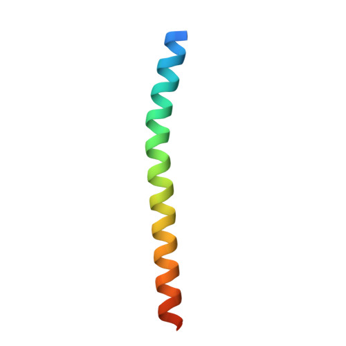
We present here the first structural report derived from breast cancer metastasis suppressor 1 (BRMS1), a member of the metastasis suppressor protein group, which, during recent years, have drawn much attention since they suppress metastasis without affecting the growth of the primary tumor. The relevance of the predicted N-terminal coiled coil on the molecular recognition of some of the BRMS1 partners, on its cellular localization and on the role of BRMS1 biological functions such as transcriptional repression prompted us to characterize its three-dimensional structure by X-ray crystallography. The structure of BRMS1 N-terminal region reveals that residues 51-98 form an antiparallel coiled-coil motif and, also, that it has the capability of homo-oligomerizing in a hexameric conformation by forming a trimer of coiled-coil dimers. We have also performed hydrodynamic experiments that strongly supported the prevalence in solution of this quaternary structure for BRMS1(51-98). This work explores the structural features of BRMS1 N-terminal region to help clarify the role of this area in the context of the full-length protein. Our crystallographic and biophysical results suggest that the biological function of BRMS1 may be affected by its ability to promote molecular clustering through its N-terminal coiled-coil region.
Organizational Affiliation:
Instituto de Biomedicina de Valencia (IBV-CSIC), C/ Jaime Roig 11, 46010 Valencia, Spain.