2XSH

CRYSTAL STRUCTURE OF P4 VARIANT OF BIPHENYL DIOXYGENASE FROM BURKHOLDERIA XENOVORANS LB400 IN COMPLEX WITH 2,6 DI CHLOROBIPHENYL


Experimental Data Snapshot

  • Method: X-RAY DIFFRACTION
  • Resolution: 2.29 Å
  • R-Value Free: 0.245 
  • R-Value Work: 0.186 
  • R-Value Observed: 0.189 

wwPDB Validation   3D Report Full Report


Ligand Structure Quality Assessment 


This is version 1.3 of the entry. See complete history


Literature

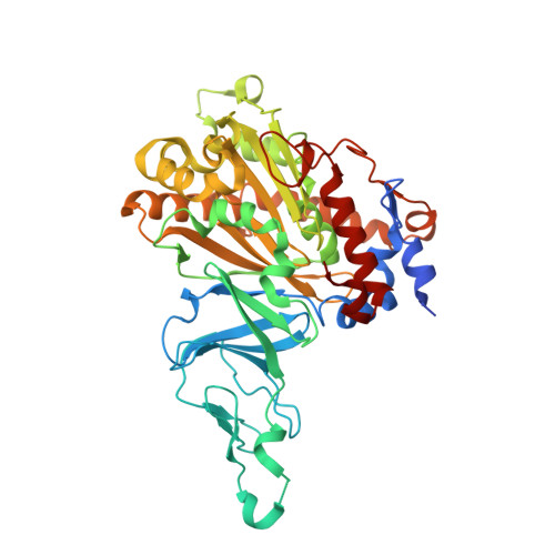
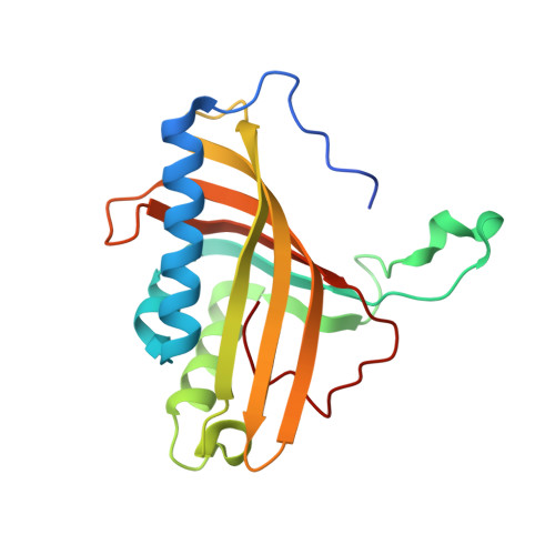
Structural Insight Into the Expanded Pcb-Degrading Abilities of a Biphenyl Dioxygenase Obtained by Directed Evolution.

Kumar, P.Mohammadi, M.Viger, J.F.Barriault, D.Gomez-Gil, L.Eltis, L.D.Bolin, J.T.Sylvestre, M.

(2011) J Mol Biol 405: 531

  • DOI: https://doi.org/10.1016/j.jmb.2010.11.009
  • Primary Citation of Related Structures:  
    2XR8, 2XRX, 2XSH, 2XSO

  • PubMed Abstract: 

    The biphenyl dioxygenase of Burkholderia xenovorans LB400 is a multicomponent Rieske-type oxygenase that catalyzes the dihydroxylation of biphenyl and many polychlorinated biphenyls (PCBs). The structural bases for the substrate specificity of the enzyme's oxygenase component (BphAE(LB400)) are largely unknown. BphAE(p4), a variant previously obtained through directed evolution, transforms several chlorobiphenyls, including 2,6-dichlorobiphenyl, more efficiently than BphAE(LB400), yet differs from the parent oxygenase at only two positions: T335A/F336M. Here, we compare the structures of BphAE(LB400) and BphAE(p4) and examine the biochemical properties of two BphAE(LB400) variants with single substitutions, T335A or F336M. Our data show that residue 336 contacts the biphenyl and influences the regiospecificity of the reaction, but does not enhance the enzyme's reactivity toward 2,6-dichlorobiphenyl. By contrast, residue 335 does not contact biphenyl but contributes significantly to expansion of the enzyme's substrate range. Crystal structures indicate that Thr335 imposes constraints through hydrogen bonds and nonbonded contacts to the segment from Val320 to Gln322. These contacts are lost when Thr is replaced by Ala, relieving intramolecular constraints and allowing for significant movement of this segment during binding of 2,6-dichlorobiphenyl, which increases the space available to accommodate the doubly ortho-chlorinated congener 2,6-dichlorobiphenyl. This study provides important insight about how Rieske-type oxygenases can expand substrate range through mutations that increase the plasticity and/or mobility of protein segments lining the catalytic cavity.


  • Organizational Affiliation

    Department of Biological Sciences, Purdue University, West Lafayette, IN 47907, USA.


Macromolecules
Find similar proteins by:  (by identity cutoff)  |  3D Structure
Entity ID: 1
MoleculeChains Sequence LengthOrganismDetailsImage
BIPHENYL DIOXYGENASE SUBUNIT ALPHA
A, C, E, G, I
A, C, E, G, I, K
459Paraburkholderia xenovorans LB400Mutation(s): 2 
EC: 1.14.12.18
UniProt
Find proteins for P37333 (Paraburkholderia xenovorans (strain LB400))
Explore P37333 
Go to UniProtKB:  P37333
Entity Groups  
Sequence Clusters30% Identity50% Identity70% Identity90% Identity95% Identity100% Identity
UniProt GroupP37333
Sequence Annotations
Expand
  • Reference Sequence
Find similar proteins by:  (by identity cutoff)  |  3D Structure
Entity ID: 2
MoleculeChains Sequence LengthOrganismDetailsImage
BIPHENYL DIOXYGENASE SUBUNIT BETA
B, D, F, H, J
B, D, F, H, J, L
188Paraburkholderia xenovorans LB400Mutation(s): 0 
EC: 1.14.12.18
UniProt
Find proteins for P37334 (Paraburkholderia xenovorans (strain LB400))
Explore P37334 
Go to UniProtKB:  P37334
Entity Groups  
Sequence Clusters30% Identity50% Identity70% Identity90% Identity95% Identity100% Identity
UniProt GroupP37334
Sequence Annotations
Expand
  • Reference Sequence
Small Molecules
Ligands 3 Unique
IDChains Name / Formula / InChI Key2D Diagram3D Interactions
DC5
Query on DC5

Download Ideal Coordinates CCD File 
O [auth A],
R [auth C],
S [auth E]
2,6-DICHLOROBIPHENYL
C12 H8 Cl2
IYZWUWBAFUBNCH-UHFFFAOYSA-N
FES
Query on FES

Download Ideal Coordinates CCD File 
M [auth A]
P [auth C]
T [auth E]
V [auth G]
X [auth I]
M [auth A],
P [auth C],
T [auth E],
V [auth G],
X [auth I],
Z [auth K]
FE2/S2 (INORGANIC) CLUSTER
Fe2 S2
NIXDOXVAJZFRNF-UHFFFAOYSA-N
FE2
Query on FE2

Download Ideal Coordinates CCD File 
AA [auth K]
N [auth A]
Q [auth C]
U [auth E]
W [auth G]
AA [auth K],
N [auth A],
Q [auth C],
U [auth E],
W [auth G],
Y [auth I]
FE (II) ION
Fe
CWYNVVGOOAEACU-UHFFFAOYSA-N
Experimental Data & Validation

Experimental Data

  • Method: X-RAY DIFFRACTION
  • Resolution: 2.29 Å
  • R-Value Free: 0.245 
  • R-Value Work: 0.186 
  • R-Value Observed: 0.189 
  • Space Group: P 1 21 1
Unit Cell:
Length ( Å )Angle ( ˚ )
a = 86.728α = 90
b = 276.762β = 117.37
c = 92.317γ = 90
Software Package:
Software NamePurpose
REFMACrefinement
HKL-2000data reduction
SCALEPACKdata scaling
MOLREPphasing

Structure Validation

View Full Validation Report


Ligand Structure Quality Assessment 


Entry History 

Deposition Data

Revision History  (Full details and data files)

  • Version 1.0: 2010-11-24
    Type: Initial release
  • Version 1.1: 2011-05-08
    Changes: Version format compliance
  • Version 1.2: 2011-07-13
    Changes: Version format compliance
  • Version 1.3: 2023-12-20
    Changes: Data collection, Database references, Derived calculations, Other, Refinement description