2XAX

Ribonucleotide reductase Y730NO2Y and Y731A modified R1 subunit of E. coli


Experimental Data Snapshot

  • Method: X-RAY DIFFRACTION
  • Resolution: 2.75 Å
  • R-Value Free: 0.261 
  • R-Value Work: 0.182 
  • R-Value Observed: 0.186 

wwPDB Validation   3D Report Full Report


This is version 1.4 of the entry. See complete history


Literature

Site-Specific Incorporation of 3-Nitrotyrosine as a Probe of Pk(A) Perturbation of Redox-Active Tyrosines in Ribonucleotide Reductase.

Yokoyama, K.Uhlin, U.Stubbe, J.

(2010) J Am Chem Soc 132: 8385

  • DOI: https://doi.org/10.1021/ja101097p
  • Primary Citation of Related Structures:  
    2X0X, 2XAK, 2XAP, 2XAV, 2XAW, 2XAX, 2XAY, 2XAZ

  • PubMed Abstract: 

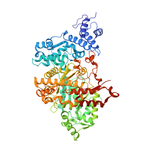
    E. coli ribonucleotide reductase catalyzes the reduction of nucleoside 5'-diphosphates into 2'-deoxynucleotides and is composed of two subunits: alpha2 and beta2. During turnover, a stable tyrosyl radical (Y*) at Y(122)-beta2 reversibly oxidizes C(439) in the active site of alpha2. This radical propagation step is proposed to occur over 35 A, to use specific redox-active tyrosines (Y(122) and Y(356) in beta2, Y(731) and Y(730) in alpha2), and to involve proton-coupled electron transfer (PCET). 3-Nitrotyrosine (NO(2)Y, pK(a) 7.1) has been incorporated in place of Y(122), Y(731), and Y(730) to probe how the protein environment perturbs each pK(a) in the presence of the second subunit, substrate (S), and allosteric effector (E). The activity of each mutant is <4 x 10(-3) that of the wild-type (wt) subunit. The [NO(2)Y(730)]-alpha2 and [NO(2)Y(731)]-alpha2 each exhibit a pK(a) of 7.8-8.0 with E and E/beta2. The pK(a) of [NO(2)Y(730)]-alpha2 is elevated to 8.2-8.3 in the S/E/beta2 complex, whereas no further perturbation is observed for [NO(2)Y(731)]-alpha2. Mutations in pathway residues adjacent to the NO(2)Y that disrupt H-bonding minimally perturb its pK(a). The pK(a) of NO(2)Y(122)-beta2 alone or with alpha2/S/E is >9.6. X-ray crystal structures have been obtained for all [NO(2)Y]-alpha2 mutants (2.1-3.1 A resolution), which show minimal structural perturbation compared to wt-alpha2. Together with the pK(a) of the previously reported NO(2)Y(356)-beta2 (7.5 in the alpha2/S/E complex; Yee, C. et al. Biochemistry 2003, 42, 14541-14552), these studies provide a picture of the protein environment of the ground state at each Y in the PCET pathway, and are the starting point for understanding differences in PCET mechanisms at each residue in the pathway.


  • Organizational Affiliation

    Department of Chemistry, Massachusetts Institute of Technology, 77 Massachusetts Avenue, Cambridge, Massachusetts 02139-4307, USA.


Macromolecules
Find similar proteins by:  (by identity cutoff)  |  3D Structure
Entity ID: 1
MoleculeChains Sequence LengthOrganismDetailsImage
RIBONUCLEOSIDE-DIPHOSPHATE REDUCTASE 1 SUBUNIT ALPHA
A, B, C
761Escherichia coli K-12Mutation(s): 1 
EC: 1.17.4.1
UniProt
Find proteins for P00452 (Escherichia coli (strain K12))
Explore P00452 
Go to UniProtKB:  P00452
Entity Groups  
Sequence Clusters30% Identity50% Identity70% Identity90% Identity95% Identity100% Identity
UniProt GroupP00452
Sequence Annotations
Expand
  • Reference Sequence

Find similar proteins by:  Sequence   |   3D Structure  

Entity ID: 2
MoleculeChains Sequence LengthOrganismDetailsImage
RIBONUCLEOSIDE-DIPHOSPHATE REDUCTASE 1 SUBUNIT BETAD,
E,
F,
G [auth P]
20synthetic constructMutation(s): 0 
EC: 1.17.4.1
UniProt
Find proteins for P69924 (Escherichia coli (strain K12))
Explore P69924 
Go to UniProtKB:  P69924
Entity Groups  
Sequence Clusters30% Identity50% Identity70% Identity90% Identity95% Identity100% Identity
UniProt GroupP69924
Sequence Annotations
Expand
  • Reference Sequence
Small Molecules
Modified Residues  1 Unique
IDChains TypeFormula2D DiagramParent
NIY
Query on NIY
A, B, C
L-PEPTIDE LINKINGC9 H10 N2 O5TYR
Experimental Data & Validation

Experimental Data

  • Method: X-RAY DIFFRACTION
  • Resolution: 2.75 Å
  • R-Value Free: 0.261 
  • R-Value Work: 0.182 
  • R-Value Observed: 0.186 
  • Space Group: H 3 2
Unit Cell:
Length ( Å )Angle ( ˚ )
a = 224.863α = 90
b = 224.863β = 90
c = 336.401γ = 120
Software Package:
Software NamePurpose
REFMACrefinement
MOSFLMdata reduction
TRUNCATEdata scaling

Structure Validation

View Full Validation Report


Entry History 

Deposition Data

Revision History  (Full details and data files)

  • Version 1.0: 2010-04-14
    Type: Initial release
  • Version 1.1: 2011-05-08
    Changes: Version format compliance
  • Version 1.2: 2011-07-13
    Changes: Version format compliance
  • Version 1.3: 2019-02-20
    Changes: Data collection, Derived calculations, Other, Source and taxonomy
  • Version 1.4: 2023-12-20
    Changes: Data collection, Database references, Other, Refinement description