The Filamentous Phages Fd and If1 Use Different Mechanisms to Infect Escherichia Coli.
Lorenz, S.H., Jakob, R.P., Weininger, U., Balbach, J., Dobbek, H., Schmid, F.X.(2011) J Mol Biol 405: 989
- PubMed: 21110981
- DOI: https://doi.org/10.1016/j.jmb.2010.11.030
- Primary Citation of Related Structures:
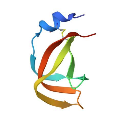
2X9A, 2X9B - PubMed Abstract:
The filamentous phage fd uses its gene 3 protein (G3P) to target Escherichia coli cells in a two-step process. First, the N2 domain of G3P attaches to an F pilus, and then the N1 domain binds to TolA-C. N1 and N2 are tightly associated, rendering the phage robust but noninfectious because the binding site for TolA-C is buried at the domain interface. Binding of N2 to the F pilus initiates partial unfolding, domain disassembly, and prolyl cis-to-trans isomerization in the hinge between N1 and N2. This activates the phage, and trans-Pro213 maintains this state long enough for N1 to reach TolA-C. Phage IF1 targets I pili, and its G3P contains also an N1 domain and an N2 domain. The pilus-binding N2 domains of the phages IF1 and fd are unrelated, and the N1 domains share a 31% sequence identity. We show that N2 of phage IF1 mediates binding to the I pilus, and that N1 targets TolA. Crystallographic and NMR analyses of the complex between N1 and TolA-C indicate that phage IF1 interacts with the same site on TolA-C as phage fd. In IF1-G3P, N1 and N2 are independently folding units, however, and the TolA binding site on N1 is permanently accessible. Activation by unfolding and prolyl isomerization, as in the case of phage fd, is not observed. In IF1-G3P, the absence of stabilizing domain interactions is compensated for by a strong increase in the stabilities of the individual domains. Apparently, these closely related filamentous phages evolved different mechanisms to reconcile robustness with high infectivity.
Organizational Affiliation:
Laboratorium für Biochemie, Universität Bayreuth, D-95440 Bayreuth, Germany.