2X7A

Structural basis of HIV-1 tethering to membranes by the Bst2-tetherin ectodomain


Experimental Data Snapshot

  • Method: X-RAY DIFFRACTION
  • Resolution: 2.77 Å
  • R-Value Free: 0.273 
  • R-Value Work: 0.236 
  • R-Value Observed: 0.238 

wwPDB Validation   3D Report Full Report


This is version 1.2 of the entry. See complete history


Literature

Structural Basis of HIV-1 Tethering to Membranes by the Bst-2/Tetherin Ectodomain.

Hinz, A.Miguet, N.Natrajan, G.Usami, Y.Yamanaka, H.Renesto, P.Hartlieb, B.Mccarthy, A.A.Simorre, J.P.Gottlinger, H.Weissenhorn, W.

(2010) Cell Host Microbe 7: 314

  • DOI: https://doi.org/10.1016/j.chom.2010.03.005
  • Primary Citation of Related Structures:  
    2X7A

  • PubMed Abstract: 

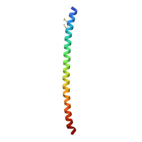
    The restriction factor BST-2/tetherin contains two membrane anchors employed to retain some enveloped viruses, including HIV-1 tethered to the plasma membrane in the absence of virus-encoded antagonists. The 2.77 A crystal structure of the BST-2/tetherin extracellular core presented here reveals a parallel 90 A long disulfide-linked coiled-coil domain, while the complete extracellular domain forms an extended 170 A long rod-like structure based on small-angle X-ray scattering data. Mutagenesis analyses indicate that both the coiled coil and the N-terminal region are required for retention of HIV-1, suggesting that the elongated structure can function as a molecular ruler to bridge long distances. The structure reveals substantial irregularities and instabilities throughout the coiled coil, which contribute to its low stability in the absence of disulfide bonds. We propose that the irregular coiled coil provides conformational flexibility, ensuring that BST-2/tetherin anchoring both in the plasma membrane and in the newly formed virus membrane is maintained during virus budding.


  • Organizational Affiliation

    Unit of Virus Host Cell Interactions (UVHCI) UMI 3265 Université Joseph Fourier-EMBL-CNRS, 6 rue Jules Horowitz, 38042 Grenoble, France.


Macromolecules
Find similar proteins by:  (by identity cutoff)  |  3D Structure
Entity ID: 1
MoleculeChains Sequence LengthOrganismDetailsImage
BONE MARROW STROMAL ANTIGEN 2
A, B, C, D, E
A, B, C, D, E, F, G, H, I, J, K
61Homo sapiensMutation(s): 0 
UniProt & NIH Common Fund Data Resources
Find proteins for Q10589 (Homo sapiens)
Explore Q10589 
Go to UniProtKB:  Q10589
PHAROS:  Q10589
GTEx:  ENSG00000130303 
Entity Groups  
Sequence Clusters30% Identity50% Identity70% Identity90% Identity95% Identity100% Identity
UniProt GroupQ10589
Sequence Annotations
Expand
  • Reference Sequence
Small Molecules
Ligands 4 Unique
IDChains Name / Formula / InChI Key2D Diagram3D Interactions
GOL
Query on GOL

Download Ideal Coordinates CCD File 
R [auth G]GLYCEROL
C3 H8 O3
PEDCQBHIVMGVHV-UHFFFAOYSA-N
CL
Query on CL

Download Ideal Coordinates CCD File 
AA [auth J]
M [auth B]
O [auth E]
Q [auth F]
S [auth G]
AA [auth J],
M [auth B],
O [auth E],
Q [auth F],
S [auth G],
T [auth G],
W [auth H]
CHLORIDE ION
Cl
VEXZGXHMUGYJMC-UHFFFAOYSA-M
MG
Query on MG

Download Ideal Coordinates CCD File 
X [auth I]MAGNESIUM ION
Mg
JLVVSXFLKOJNIY-UHFFFAOYSA-N
NA
Query on NA

Download Ideal Coordinates CCD File 
L [auth B]
N [auth D]
P [auth F]
U [auth G]
V [auth H]
L [auth B],
N [auth D],
P [auth F],
U [auth G],
V [auth H],
Y [auth I],
Z [auth J]
SODIUM ION
Na
FKNQFGJONOIPTF-UHFFFAOYSA-N
Modified Residues  1 Unique
IDChains TypeFormula2D DiagramParent
MSE
Query on MSE
A, B, C, D, E
A, B, C, D, E, F, G, H, I, J, K
L-PEPTIDE LINKINGC5 H11 N O2 SeMET
Experimental Data & Validation

Experimental Data

  • Method: X-RAY DIFFRACTION
  • Resolution: 2.77 Å
  • R-Value Free: 0.273 
  • R-Value Work: 0.236 
  • R-Value Observed: 0.238 
  • Space Group: C 1 2 1
Unit Cell:
Length ( Å )Angle ( ˚ )
a = 169.89α = 90
b = 85.93β = 126.94
c = 123.31γ = 90
Software Package:
Software NamePurpose
PHENIXrefinement
XDSdata reduction
SCALAdata scaling
SHARPphasing

Structure Validation

View Full Validation Report


Entry History 

Deposition Data

Revision History  (Full details and data files)

  • Version 1.0: 2010-04-28
    Type: Initial release
  • Version 1.1: 2011-06-02
    Changes: Version format compliance
  • Version 1.2: 2011-07-13
    Changes: Version format compliance