2X0R

R207S, R292S Mutant of Malate Dehydrogenase from the Halophilic Archeon Haloarcula marismortui (HoloForm)


Experimental Data Snapshot

  • Method: X-RAY DIFFRACTION
  • Resolution: 2.92 Å
  • R-Value Free: 0.286 
  • R-Value Work: 0.184 
  • R-Value Observed: 0.189 

wwPDB Validation   3D Report Full Report


Ligand Structure Quality Assessment 


This is version 1.4 of the entry. See complete history


Literature

The Oligomeric States of Haloarcula Marismortui Malate Dehydrogenase are Modulated by Solvent Components as Shown by Crystallographic and Biochemical Studies

Irimia, A.Ebel, C.Madern, D.Richard, S.B.Cosenza, L.W.Zaccai, G.Vellieux, F.M.D.

(2003) J Mol Biol 326: 859-873

  • DOI: https://doi.org/10.1016/s0022-2836(02)01450-x
  • Primary Citation of Related Structures:  
    1O6Z, 2X0R

  • PubMed Abstract: 

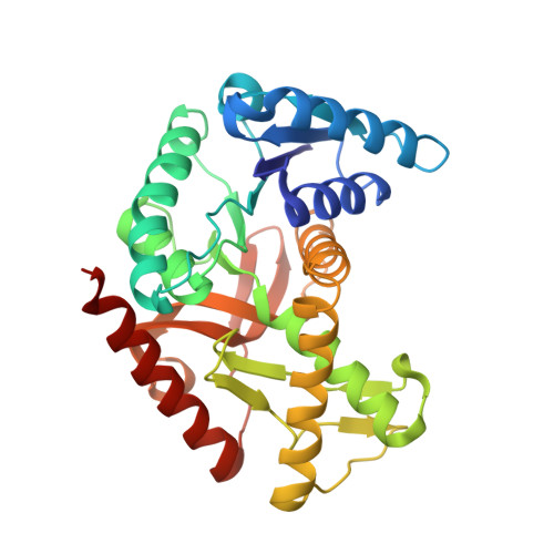
    The three-dimensional crystal structure of the (R207S, R292S) mutant of malate dehydrogenase from Haloarcula marismortui was solved at 1.95A resolution in order to determine the role of salt bridges and solvent ions in halophilic adaptation and quaternary structure stability. The mutations, located at the dimer-dimer interface, disrupt two inter-dimeric salt bridge clusters that are essential for wild-type tetramer stabilisation. Previous experiments in solution, performed on the double mutant, had shown a tetrameric structure in 4M NaCl, which dissociated into active dimers in 2M NaCl. In order to establish if the active dimeric form is a product of the mutation, or if it also exists in the wild-type protein, complementary studies were performed on the wild-type enzyme by analytical centrifugation and small angle neutron scattering experiments. They showed the existence of active dimers in NaF, KF, Na(2)SO(4), even in the absence of NADH, and in the presence of NADH at concentrations of NaCl below 0.3M. The crystal structure shows a tetramer that, in the absence of the salt bridge clusters, appears to be stabilized by a network of ordered water molecules and by Cl(-) binding at the dimer-dimer interface. The double mutant and wild-type dimer folds are essentially identical (the r.m.s. deviation between equivalent C(alpha) positions is 0.39A). Chloride ions are also observed at the monomer-monomer interfaces of the mutant, contributing to the stability of each dimer against low salt dissociation. Our results support the hypothesis that extensive binding of water and salt is an important feature of adaptation to a halophilic environment.


  • Organizational Affiliation

    Laboratoire de Biophysique Moléculaire, Institut de Biologie Structurale J.-P. Ebel CEA CNRS UJF UMR-5075, 41 rue Jules Horowitz, 38027 Grenoble Cedex 01, France.


Macromolecules
Find similar proteins by:  (by identity cutoff)  |  3D Structure
Entity ID: 1
MoleculeChains Sequence LengthOrganismDetailsImage
MALATE DEHYDROGENASE
A, B
304Haloarcula marismortuiMutation(s): 2 
EC: 1.1.1.37
UniProt
Find proteins for Q07841 (Haloarcula marismortui (strain ATCC 43049 / DSM 3752 / JCM 8966 / VKM B-1809))
Explore Q07841 
Go to UniProtKB:  Q07841
Entity Groups  
Sequence Clusters30% Identity50% Identity70% Identity90% Identity95% Identity100% Identity
UniProt GroupQ07841
Sequence Annotations
Expand
  • Reference Sequence
Small Molecules
Ligands 3 Unique
IDChains Name / Formula / InChI Key2D Diagram3D Interactions
NAD
Query on NAD

Download Ideal Coordinates CCD File 
C [auth A],
G [auth B]
NICOTINAMIDE-ADENINE-DINUCLEOTIDE
C21 H27 N7 O14 P2
BAWFJGJZGIEFAR-NNYOXOHSSA-N
CL
Query on CL

Download Ideal Coordinates CCD File 
D [auth A],
E [auth A],
H [auth B],
I [auth B]
CHLORIDE ION
Cl
VEXZGXHMUGYJMC-UHFFFAOYSA-M
NA
Query on NA

Download Ideal Coordinates CCD File 
F [auth A],
J [auth B],
K [auth B],
L [auth B],
M [auth B]
SODIUM ION
Na
FKNQFGJONOIPTF-UHFFFAOYSA-N
Experimental Data & Validation

Experimental Data

  • Method: X-RAY DIFFRACTION
  • Resolution: 2.92 Å
  • R-Value Free: 0.286 
  • R-Value Work: 0.184 
  • R-Value Observed: 0.189 
  • Space Group: C 2 2 21
Unit Cell:
Length ( Å )Angle ( ˚ )
a = 115.36α = 90
b = 125.91β = 90
c = 125.84γ = 90
Software Package:
Software NamePurpose
PHENIXrefinement
XDSdata reduction
BIOMOLdata scaling
AMoREphasing

Structure Validation

View Full Validation Report


Ligand Structure Quality Assessment 


Entry History 

Deposition Data

Revision History  (Full details and data files)

  • Version 1.0: 2009-12-29
    Type: Initial release
  • Version 1.1: 2011-05-08
    Changes: Version format compliance
  • Version 1.2: 2011-07-13
    Changes: Version format compliance
  • Version 1.3: 2018-06-13
    Changes: Data collection, Database references, Structure summary
  • Version 1.4: 2023-12-20
    Changes: Data collection, Database references, Derived calculations, Other, Refinement description