2WZG

Legionella glucosyltransferase (Lgt1) crystal structure


Experimental Data Snapshot

  • Method: X-RAY DIFFRACTION
  • Resolution: 1.90 Å
  • R-Value Free: 0.262 
  • R-Value Work: 0.200 
  • R-Value Observed: 0.201 

wwPDB Validation   3D Report Full Report


Ligand Structure Quality Assessment 


This is version 1.3 of the entry. See complete history


Literature

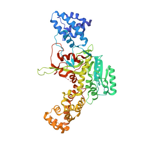
Molecular Mechanism of Elongation Factor 1A Inhibition by a Legionella Pneumophila Glycosyltransferase.

Hurtado-Guerrero, R.Zusman, T.Pathak, S.Ibrahim, A.F.M.Shepherd, S.Prescott, A.Segal, G.Van Aalten, D.M.F.

(2010) Biochem J 426: 281

  • DOI: https://doi.org/10.1042/BJ20091351
  • Primary Citation of Related Structures:  
    2WZF, 2WZG

  • PubMed Abstract: 

    Legionnaires' disease is caused by a lethal colonization of alveolar macrophages with the Gram-negative bacterium Legionella pneumophila. LpGT (L. pneumophila glucosyltransferase; also known as Lgt1) has recently been identified as a virulence factor, shutting down protein synthesis in the human cell by specific glucosylation of EF1A (elongation factor 1A), using an unknown mode of substrate recognition and a retaining mechanism for glycosyl transfer. We have determined the crystal structure of LpGT in complex with substrates, revealing a GT-A fold with two unusual protruding domains. Through structure-guided mutagenesis of LpGT, several residues essential for binding of the UDP-glucose-donor and EF1A-acceptor substrates were identified, which also affected L. pneumophila virulence as demonstrated by microinjection studies. Together, these results suggested that a positively charged EF1A loop binds to a negatively charged conserved groove on the LpGT structure, and that two asparagine residues are essential for catalysis. Furthermore, we showed that two further L. pneumophila glycosyltransferases possessed the conserved UDP-glucose-binding sites and EF1A-binding grooves, and are, like LpGT, translocated into the macrophage through the Icm/Dot (intracellular multiplication/defect in organelle trafficking) system.


  • Organizational Affiliation

    Division of Molecular Microbiology, College of Life Sciences, University of Dundee, Dundee DD1 5EH, Scotland, UK. R.HurtadoGuerrero@dundee.ac.uk


Macromolecules
Find similar proteins by:  (by identity cutoff)  |  3D Structure
Entity ID: 1
MoleculeChains Sequence LengthOrganismDetailsImage
GLUCOSYLTRANSFERASE525Legionella pneumophilaMutation(s): 0 
UniProt
Find proteins for Q5WWY0 (Legionella pneumophila (strain Lens))
Explore Q5WWY0 
Go to UniProtKB:  Q5WWY0
Entity Groups  
Sequence Clusters30% Identity50% Identity70% Identity90% Identity95% Identity100% Identity
UniProt GroupQ5WWY0
Sequence Annotations
Expand
  • Reference Sequence
Small Molecules
Ligands 2 Unique
IDChains Name / Formula / InChI Key2D Diagram3D Interactions
UPG
Query on UPG

Download Ideal Coordinates CCD File 
C [auth A]URIDINE-5'-DIPHOSPHATE-GLUCOSE
C15 H24 N2 O17 P2
HSCJRCZFDFQWRP-JZMIEXBBSA-N
MG
Query on MG

Download Ideal Coordinates CCD File 
B [auth A]MAGNESIUM ION
Mg
JLVVSXFLKOJNIY-UHFFFAOYSA-N
Binding Affinity Annotations 
IDSourceBinding Affinity
UPG Binding MOAD:  2WZG Kd: 100 (nM) from 1 assay(s)
Experimental Data & Validation

Experimental Data

  • Method: X-RAY DIFFRACTION
  • Resolution: 1.90 Å
  • R-Value Free: 0.262 
  • R-Value Work: 0.200 
  • R-Value Observed: 0.201 
  • Space Group: H 3
Unit Cell:
Length ( Å )Angle ( ˚ )
a = 122.75α = 90
b = 122.75β = 90
c = 103.416γ = 120
Software Package:
Software NamePurpose
REFMACrefinement
DENZOdata reduction
SCALEPACKdata scaling
SHELXphasing

Structure Validation

View Full Validation Report


Ligand Structure Quality Assessment 


Entry History 

Deposition Data

Revision History  (Full details and data files)

  • Version 1.0: 2009-12-08
    Type: Initial release
  • Version 1.1: 2011-05-08
    Changes: Version format compliance
  • Version 1.2: 2011-07-13
    Changes: Version format compliance
  • Version 1.3: 2018-05-02
    Changes: Data collection, Source and taxonomy, Structure summary