2WYH

Structure of the Streptococcus pyogenes family GH38 alpha-mannosidase


Experimental Data Snapshot

  • Method: X-RAY DIFFRACTION
  • Resolution: 1.90 Å
  • R-Value Free: 0.205 
  • R-Value Work: 0.177 
  • R-Value Observed: 0.179 

wwPDB Validation   3D Report Full Report


This is version 1.1 of the entry. See complete history


Literature

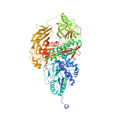
Structure and Kinetic Investigation of Streptococcus Pyogenes Family Gh38 Alpha-Mannosidase

Suits, M.D.L.Zhu, Y.Taylor, E.J.Zechel, D.L.Gilbert, H.J.Davies, G.J.

(2010) PLoS One 5: E9006

  • DOI: https://doi.org/10.1371/journal.pone.0009006
  • Primary Citation of Related Structures:  
    2WYH, 2WYI

  • PubMed Abstract: 

    The enzymatic hydrolysis of alpha-mannosides is catalyzed by glycoside hydrolases (GH), termed alpha-mannosidases. These enzymes are found in different GH sequence-based families. Considerable research has probed the role of higher eukaryotic "GH38" alpha-mannosides that play a key role in the modification and diversification of hybrid N-glycans; processes with strong cellular links to cancer and autoimmune disease. The most extensively studied of these enzymes is the Drosophila GH38 alpha-mannosidase II, which has been shown to be a retaining alpha-mannosidase that targets both alpha-1,3 and alpha-1,6 mannosyl linkages, an activity that enables the enzyme to process GlcNAc(Man)(5)(GlcNAc)(2) hybrid N-glycans to GlcNAc(Man)(3)(GlcNAc)(2). Far less well understood is the observation that many bacterial species, predominantly but not exclusively pathogens and symbionts, also possess putative GH38 alpha-mannosidases whose activity and specificity is unknown.


  • Organizational Affiliation

    York Structural Biology Laboratory, Department of Chemistry, University of York, York, United Kingdom.


Macromolecules
Find similar proteins by:  (by identity cutoff)  |  3D Structure
Entity ID: 1
MoleculeChains Sequence LengthOrganismDetailsImage
ALPHA-MANNOSIDASE
A, B
923Streptococcus pyogenes M1 GASMutation(s): 0 
EC: 3.2.1.24
UniProt
Find proteins for Q99YP5 (Streptococcus pyogenes serotype M1)
Explore Q99YP5 
Go to UniProtKB:  Q99YP5
Entity Groups  
Sequence Clusters30% Identity50% Identity70% Identity90% Identity95% Identity100% Identity
UniProt GroupQ99YP5
Sequence Annotations
Expand
  • Reference Sequence
Small Molecules
Ligands 3 Unique
IDChains Name / Formula / InChI Key2D Diagram3D Interactions
TRS
Query on TRS

Download Ideal Coordinates CCD File 
L [auth B]2-AMINO-2-HYDROXYMETHYL-PROPANE-1,3-DIOL
C4 H12 N O3
LENZDBCJOHFCAS-UHFFFAOYSA-O
GOL
Query on GOL

Download Ideal Coordinates CCD File 
D [auth A]
E [auth A]
F [auth A]
G [auth A]
H [auth A]
D [auth A],
E [auth A],
F [auth A],
G [auth A],
H [auth A],
I [auth A],
J [auth A],
M [auth B],
N [auth B],
O [auth B],
P [auth B],
Q [auth B]
GLYCEROL
C3 H8 O3
PEDCQBHIVMGVHV-UHFFFAOYSA-N
ZN
Query on ZN

Download Ideal Coordinates CCD File 
C [auth A],
K [auth B]
ZINC ION
Zn
PTFCDOFLOPIGGS-UHFFFAOYSA-N
Experimental Data & Validation

Experimental Data

  • Method: X-RAY DIFFRACTION
  • Resolution: 1.90 Å
  • R-Value Free: 0.205 
  • R-Value Work: 0.177 
  • R-Value Observed: 0.179 
  • Space Group: P 1 21 1
Unit Cell:
Length ( Å )Angle ( ˚ )
a = 92.573α = 90
b = 88.502β = 108.97
c = 134.688γ = 90
Software Package:
Software NamePurpose
REFMACrefinement
HKL-2000data reduction
SCALEPACKdata scaling
SHARPphasing
SHELXphasing
RESOLVEphasing

Structure Validation

View Full Validation Report


Entry History 

Deposition Data

Revision History  (Full details and data files)

  • Version 1.0: 2010-02-16
    Type: Initial release
  • Version 1.1: 2011-07-13
    Changes: Advisory, Refinement description, Version format compliance