2WL7

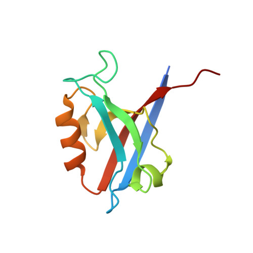
Crystal structure of the PSD93 PDZ1 domain


Experimental Data Snapshot

  • Method: X-RAY DIFFRACTION
  • Resolution: 2.03 Å
  • R-Value Free: 0.208 
  • R-Value Work: 0.164 
  • R-Value Observed: 0.169 

wwPDB Validation   3D Report Full Report


This is version 1.3 of the entry. See complete history


Literature

Structure of the First Pdz Domain of Human Psd-93.

Fiorentini, M.Nielsen, A.K.Kristensen, O.Kastrup, J.S.Gajhede, M.

(2009) Acta Crystallogr Sect F Struct Biol Cryst Commun 65: 1254

  • DOI: https://doi.org/10.1107/S1744309109043267
  • Primary Citation of Related Structures:  
    2WL7

  • PubMed Abstract: 

    The crystal structure of the PDZ1 domain of human PSD-93 has been determined to 2.0 A resolution. The PDZ1 domain forms a crystallographic trimer that is also predicted to be stable in solution. The main contributions to the stabilization of the trimer seem to arise from interactions involving the PDZ1-PDZ2 linker region at the extreme C-terminus of PDZ1, implying that the oligomerization that is observed is not of biological significance in full-length PSD-93. Comparison of the structures of the binding cleft of PSD-93 PDZ1 with the previously reported structures of PSD-93 PDZ2 and PDZ3 as well as of the closely related human PSD-95 PDZ1 shows that they are very similar in terms of amino-acid composition. However, the cleft is significantly narrower in PSD-95. This could be part of the basis of peptide selectivity between PSD-93 PDZ1 and PSD-95 PDZ1.


  • Organizational Affiliation

    Biostructural Research, Department of Medicinal Chemistry, Faculty of Pharmaceutical Sciences, University of Copenhagen, Universitetsparken 2, DK-2100 Copenhagen, Denmark.


Macromolecules
Find similar proteins by:  (by identity cutoff)  |  3D Structure
Entity ID: 1
MoleculeChains Sequence LengthOrganismDetailsImage
DISKS LARGE HOMOLOG 2102Mus musculusMutation(s): 0 
EC: 2.7.4.8
UniProt & NIH Common Fund Data Resources
Find proteins for Q91XM9 (Mus musculus)
Explore Q91XM9 
Go to UniProtKB:  Q91XM9
IMPC:  MGI:1344351
Entity Groups  
Sequence Clusters30% Identity50% Identity70% Identity90% Identity95% Identity100% Identity
UniProt GroupQ91XM9
Sequence Annotations
Expand
  • Reference Sequence
Experimental Data & Validation

Experimental Data

  • Method: X-RAY DIFFRACTION
  • Resolution: 2.03 Å
  • R-Value Free: 0.208 
  • R-Value Work: 0.164 
  • R-Value Observed: 0.169 
  • Space Group: H 3
Unit Cell:
Length ( Å )Angle ( ˚ )
a = 85.198α = 90
b = 85.198β = 90
c = 45.515γ = 120
Software Package:
Software NamePurpose
PHENIXrefinement
MOSFLMdata reduction
SCALAdata scaling
PHASERphasing

Structure Validation

View Full Validation Report


Entry History 

Deposition Data

Revision History  (Full details and data files)

  • Version 1.0: 2010-01-19
    Type: Initial release
  • Version 1.1: 2011-05-08
    Changes: Version format compliance
  • Version 1.2: 2011-07-13
    Changes: Version format compliance
  • Version 1.3: 2023-12-13
    Changes: Data collection, Database references, Derived calculations, Other, Refinement description