2WGZ

Crystal structure of alpha-1,3 galactosyltransferase (alpha3GT) in a complex with p-nitrophenyl-beta-galactoside (pNP-beta-Gal)


Experimental Data Snapshot

  • Method: X-RAY DIFFRACTION
  • Resolution: 2.12 Å
  • R-Value Free: 0.206 
  • R-Value Work: 0.169 
  • R-Value Observed: 0.170 

wwPDB Validation   3D Report Full Report


Ligand Structure Quality Assessment 


This is version 2.1 of the entry. See complete history


Literature

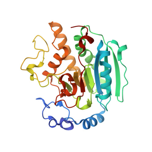
Crystal Structure of Alpha-1,3 Galactosyltransferase (Alpha3Gt) in a Complex with P-Nitrophenyl-Beta-Galactoside (Pnpbetagal).

Jamaluddin, H.Tumbale, P.Ferns, T.A.Thiyagarajan, N.Brew, K.Acharya, K.R.

(2009) Biochem Biophys Res Commun 385: 601

  • DOI: https://doi.org/10.1016/j.bbrc.2009.05.111
  • Primary Citation of Related Structures:  
    2WGZ

  • PubMed Abstract: 

    The specificities of glycosyltransferases make them useful for the synthesis of biologically active oligosaccharides, but also restrict their range of products. In substrate engineering, substrate promiscuity is enhanced by attaching removable interactive groups to weak substrates. Thus, the attachment of betap-nitrophenyl converts galactose from a poor into a good substrate of alpha-1,3-galactosyltransferase. The crystallographic structure of a complex of alpha3GT containing p-nitrophenyl-beta-galactoside shows that the p-nitrophenyl binds similarly to the N-acetylglucosamine of the substrate, N-acetyllactosamine, interacting with the indole of Trp249. p-Nitrophenyl, unlike N-acetylglucosamine, makes no H-bonds but has more non-polar interactions, making it an effective monosaccharide mimetic.


  • Organizational Affiliation

    Department of Biology and Biochemistry, University of Bath, Claverton Down, Bath BA27AY, UK.


Macromolecules
Find similar proteins by:  (by identity cutoff)  |  3D Structure
Entity ID: 1
MoleculeChains Sequence LengthOrganismDetailsImage
N-ACETYLLACTOSAMINIDE ALPHA-1,3-GALACTOSYL TRANSFERASE
A, B
290Bos taurusMutation(s): 0 
EC: 2.4.1.151 (PDB Primary Data), 2.4.1.87 (PDB Primary Data)
UniProt
Find proteins for P14769 (Bos taurus)
Explore P14769 
Go to UniProtKB:  P14769
Entity Groups  
Sequence Clusters30% Identity50% Identity70% Identity90% Identity95% Identity100% Identity
UniProt GroupP14769
Sequence Annotations
Expand
  • Reference Sequence
Small Molecules
Ligands 6 Unique
IDChains Name / Formula / InChI Key2D Diagram3D Interactions
UDP
Query on UDP

Download Ideal Coordinates CCD File 
E [auth A],
M [auth B]
URIDINE-5'-DIPHOSPHATE
C9 H14 N2 O12 P2
XCCTYIAWTASOJW-XVFCMESISA-N
147
Query on 147

Download Ideal Coordinates CCD File 
C [auth A]4-nitrophenyl beta-D-galactopyranoside
C12 H15 N O8
IFBHRQDFSNCLOZ-YBXAARCKSA-N
GAL
Query on GAL

Download Ideal Coordinates CCD File 
J [auth B]beta-D-galactopyranose
C6 H12 O6
WQZGKKKJIJFFOK-FPRJBGLDSA-N
NPO
Query on NPO

Download Ideal Coordinates CCD File 
K [auth B]P-NITROPHENOL
C6 H5 N O3
BTJIUGUIPKRLHP-UHFFFAOYSA-N
GOL
Query on GOL

Download Ideal Coordinates CCD File 
F [auth A]
G [auth A]
H [auth A]
I [auth A]
N [auth B]
F [auth A],
G [auth A],
H [auth A],
I [auth A],
N [auth B],
O [auth B]
GLYCEROL
C3 H8 O3
PEDCQBHIVMGVHV-UHFFFAOYSA-N
MN
Query on MN

Download Ideal Coordinates CCD File 
D [auth A],
L [auth B]
MANGANESE (II) ION
Mn
WAEMQWOKJMHJLA-UHFFFAOYSA-N
Experimental Data & Validation

Experimental Data

  • Method: X-RAY DIFFRACTION
  • Resolution: 2.12 Å
  • R-Value Free: 0.206 
  • R-Value Work: 0.169 
  • R-Value Observed: 0.170 
  • Space Group: P 1 21 1
Unit Cell:
Length ( Å )Angle ( ˚ )
a = 45.146α = 90
b = 94.222β = 99.05
c = 94.465γ = 90
Software Package:
Software NamePurpose
REFMACrefinement
DENZOdata reduction
SCALEPACKdata scaling
PHASERphasing

Structure Validation

View Full Validation Report


Ligand Structure Quality Assessment 


Entry History 

Deposition Data

Revision History  (Full details and data files)

  • Version 1.0: 2009-06-16
    Type: Initial release
  • Version 1.1: 2011-06-30
    Changes: Version format compliance
  • Version 1.2: 2011-07-13
    Changes: Version format compliance
  • Version 2.0: 2020-07-29
    Type: Remediation
    Reason: Carbohydrate remediation
    Changes: Atomic model, Data collection, Derived calculations, Other, Structure summary
  • Version 2.1: 2023-12-13
    Changes: Data collection, Database references, Refinement description, Structure summary