Structural Comparison of Two Cspg-Binding Dbl Domains from the Var2Csa Protein Important in Malaria During Pregnancy.
Khunrae, P., Philip, J.M.D., Bull, D.R., Higgins, M.K.(2009) J Mol Biol 393: 202
- PubMed: 19695262
- DOI: https://doi.org/10.1016/j.jmb.2009.08.027
- Primary Citation of Related Structures:
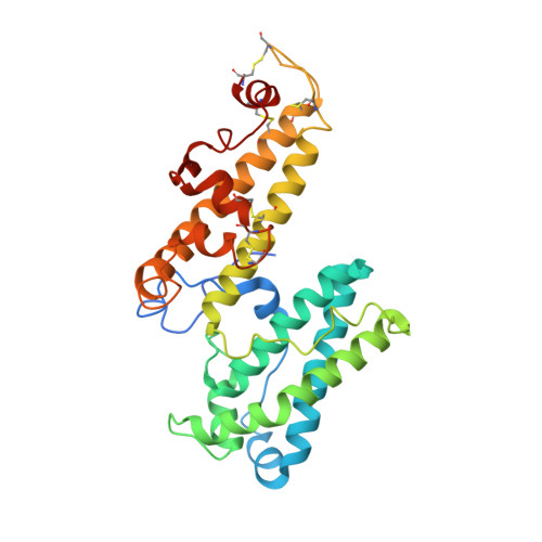
2WAU - PubMed Abstract:
Severe malaria during pregnancy is associated with accumulation of parasite-infected erythrocytes in the placenta due to interactions between VAR2CSA protein, expressed on the surface of infected-erythrocytes, and placental chondroitin sulfate proteoglycans (CSPG). VAR2CSA contains multiple CSPG-binding domains, including DBL3X and DBL6 epsilon. Previous structural studies of DBL3X suggested CSPG to bind to a positively charged patch and sulfate-binding site on the concave surface of the domain. Here we present the structure of the DBL6 epsilon domain from VAR2CSA. This domain displays the same overall architecture and secondary structure as that of DBL3X but differs in loop structures, disulfide bond positions and surface charge distribution. In particular, despite binding to CSPG, DBL6 epsilon lacks the key features of the CSPG-binding site of DBL3X. Instead DBL6 epsilon binds to CSPG through a positively charged surface on the distal side of subdomain 2 that is exposed in intact VAR2CSA on the erythrocyte surface. Finally, unlike intact VAR2CSA, both DBL3X and DBL6 epsilon bind to various carbohydrates, with greatest affinity for ligands with high sulfation and negative charge. These studies provide further insight into the structure of DBL domains and suggest a model for the role of individual domains in CSPG binding by VAR2CSA in placental malaria.
Organizational Affiliation:
Department of Biochemistry, University of Cambridge, 80, Tennis Court Road, Cambridge CB2 1GA, UK.