2W95

STructure of the Discoidin I from Dictyostelium discoideum in complex with GalNAc at 1.75 angstrom resolution


Experimental Data Snapshot

  • Method: X-RAY DIFFRACTION
  • Resolution: 1.75 Å
  • R-Value Free: 0.193 
  • R-Value Work: 0.157 
  • R-Value Observed: 0.159 

wwPDB Validation   3D Report Full Report


Ligand Structure Quality Assessment 


This is version 1.4 of the entry. See complete history


Literature

Discoidin I from Dictyostelium Discoideum and Interactions with Oligosaccharides: Specificity, Affinity, Crystal Structures and Comparison with Discoidin II.

Mathieu, S.Saboia Aragao, K.Imberty, A.Varrot, A.

(2010) J Mol Biol 400: 540

  • DOI: https://doi.org/10.1016/j.jmb.2010.05.042
  • Primary Citation of Related Structures:  
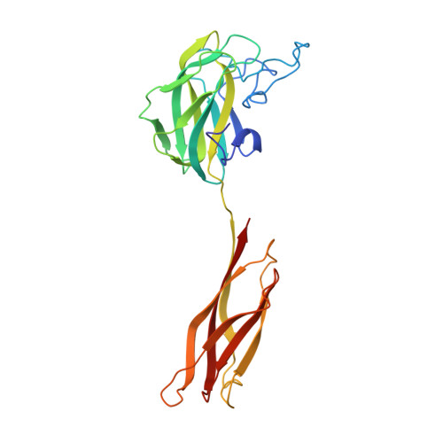
    2W94, 2W95, 2WN2, 2WN3

  • PubMed Abstract: 

    Discoidin I (DiscI) and discoidin II (DiscII) are N-acetylgalactosamine (GalNAc)-binding proteins from Dictyostelium discoideum. They consist of two domains: an N-terminal discoidin domain and a C-terminal H-type lectin domain. They were cloned and expressed in high yield in recombinant form in Escherichia coli. Although both lectins bind galactose (Gal) and GalNAc, glycan array experiments performed on the recombinant proteins displayed strong differences in their specificity for oligosaccharides. DiscI and DiscII bind preferentially to Gal/GalNAcbeta1-3Gal/GalNAc-containing and Gal/GalNAcbeta1-4GlcNAcbeta1-6Gal/GalNAc-containing glycans, respectively. The affinity of the interaction of DiscI with monosaccharides and disaccharides was evaluated using isothermal titration calorimetry experiments. The three-dimensional structures of native DiscI and its complexes with GalNAc, GalNAcbeta1-3Gal, and Galbeta1-3GalNAc were solved by X-ray crystallography. DiscI forms trimers with involvement of calcium at the monomer interface. The N-terminal discoidin domain presents a structural similarity to F-type lectins such as the eel agglutinin, where an amphiphilic binding pocket suggests possible carbohydrate-binding activity. In the C-terminal H-type lectin domain, the GalNAc residue establishes specific hydrogen bonds that explain the observed affinity (K(d)=3x10(-4) M). The different specificities of DiscI and DiscII for oligosaccharides were rationalized from the different structures obtained by either X-ray crystallography or molecular modeling.


  • Organizational Affiliation

    CERMAV-CNRS, 601 rue de la Chimie, BP53, F-38041 Grenoble Cedex 09, France.


Macromolecules
Find similar proteins by:  (by identity cutoff)  |  3D Structure
Entity ID: 1
MoleculeChains Sequence LengthOrganismDetailsImage
DISCOIDIN-1 SUBUNIT A
A, B, C
254Dictyostelium discoideum AX2Mutation(s): 0 
UniProt
Find proteins for P02886 (Dictyostelium discoideum)
Explore P02886 
Go to UniProtKB:  P02886
Entity Groups  
Sequence Clusters30% Identity50% Identity70% Identity90% Identity95% Identity100% Identity
UniProt GroupP02886
Sequence Annotations
Expand
  • Reference Sequence
Small Molecules
Ligands 6 Unique
IDChains Name / Formula / InChI Key2D Diagram3D Interactions
NGA
Query on NGA

Download Ideal Coordinates CCD File 
D [auth A],
J [auth B],
R [auth C]
2-acetamido-2-deoxy-beta-D-galactopyranose
C8 H15 N O6
OVRNDRQMDRJTHS-JAJWTYFOSA-N
MRD
Query on MRD

Download Ideal Coordinates CCD File 
N [auth B],
S [auth C],
V [auth C]
(4R)-2-METHYLPENTANE-2,4-DIOL
C6 H14 O2
SVTBMSDMJJWYQN-RXMQYKEDSA-N
MPD
Query on MPD

Download Ideal Coordinates CCD File 
E [auth A],
G [auth A],
L [auth B],
T [auth C]
(4S)-2-METHYL-2,4-PENTANEDIOL
C6 H14 O2
SVTBMSDMJJWYQN-YFKPBYRVSA-N
SO4
Query on SO4

Download Ideal Coordinates CCD File 
F [auth A]
H [auth A]
K [auth B]
M [auth B]
O [auth B]
F [auth A],
H [auth A],
K [auth B],
M [auth B],
O [auth B],
U [auth C],
W [auth C]
SULFATE ION
O4 S
QAOWNCQODCNURD-UHFFFAOYSA-L
NI
Query on NI

Download Ideal Coordinates CCD File 
Q [auth B]NICKEL (II) ION
Ni
VEQPNABPJHWNSG-UHFFFAOYSA-N
CA
Query on CA

Download Ideal Coordinates CCD File 
I [auth A],
P [auth B],
X [auth C]
CALCIUM ION
Ca
BHPQYMZQTOCNFJ-UHFFFAOYSA-N
Binding Affinity Annotations 
IDSourceBinding Affinity
NGA Binding MOAD:  2W95 Kd: 3.02e+5 (nM) from 1 assay(s)
Experimental Data & Validation

Experimental Data

  • Method: X-RAY DIFFRACTION
  • Resolution: 1.75 Å
  • R-Value Free: 0.193 
  • R-Value Work: 0.157 
  • R-Value Observed: 0.159 
  • Space Group: C 2 2 21
Unit Cell:
Length ( Å )Angle ( ˚ )
a = 72.404α = 90
b = 274.047β = 90
c = 86.136γ = 90
Software Package:
Software NamePurpose
REFMACrefinement
MOSFLMdata reduction
SCALAdata scaling
PHASERphasing

Structure Validation

View Full Validation Report


Ligand Structure Quality Assessment 


Entry History 

Deposition Data

Revision History  (Full details and data files)

  • Version 1.0: 2010-03-31
    Type: Initial release
  • Version 1.1: 2011-05-08
    Changes: Version format compliance
  • Version 1.2: 2011-07-13
    Changes: Version format compliance
  • Version 1.3: 2020-07-29
    Type: Remediation
    Reason: Carbohydrate remediation
    Changes: Data collection, Derived calculations, Other, Structure summary
  • Version 1.4: 2023-12-13
    Changes: Data collection, Database references, Refinement description, Structure summary