2VXJ

CRYSTAL STRUCTURE OF PA-IL LECTIN COMPLEXED WITH AGAL13BGAL14GLC AT 1.9 A RESOLUTION


Experimental Data Snapshot

  • Method: X-RAY DIFFRACTION
  • Resolution: 1.90 Å
  • R-Value Free: 0.245 
  • R-Value Work: 0.185 
  • R-Value Observed: 0.188 

wwPDB Validation   3D Report Full Report


Ligand Structure Quality Assessment 


This is version 2.1 of the entry. See complete history


Literature

Structural Basis of the Preferential Binding for Globo-Series Glycosphingolipids Displayed by Pseudomonas Aeruginosa Lectin I.

Blanchard, B.Nurisso, A.Hollville, E.Tetaud, C.Wiels, J.Pokorna, M.Wimmerova, M.Varrot, A.Imberty, A.

(2008) J Mol Biol 383: 837

  • DOI: https://doi.org/10.1016/j.jmb.2008.08.028
  • Primary Citation of Related Structures:  
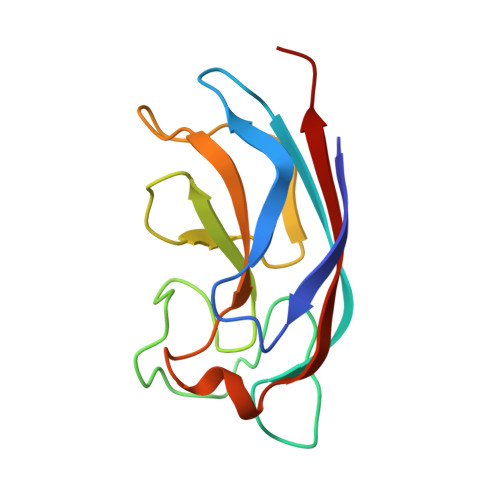
    2VXJ

  • PubMed Abstract: 

    The opportunistic pathogen Pseudomonas aeruginosa contains several carbohydrate-binding proteins, among which is the P. aeruginosa lectin I (PA-IL), which displays affinity for alpha-galactosylated glycans. Glycan arrays were screened and demonstrated stronger binding of PA-IL toward alphaGal1-4betaGal-terminating structures and weaker binding to alphaGal1-3betaGal ones in order to determine which human glycoconjugates could play a role in the carbohydrate-mediated adhesion of the bacteria. This was confirmed in vivo by testing the binding of the lectin to Burkitt lymphoma cells that present large amounts of globotriaosylceramide antigen Gb3/CD77/P(k). Trisaccharide moieties of Gb3 (alphaGal1-4betaGal1-4Glc) and isoglobotriaosylceramide (alphaGal1-3betaGal1-4Glc) were tested by titration microcalorimetry, and both displayed similar affinity to PA-IL in solution. The crystal structure of PA-IL complexed to alphaGal1-3betaGal1-4Glc trisaccharide has been solved at 1.9-A resolution and revealed how the second galactose residue makes specific contacts with the protein surface. Molecular modeling studies were performed in order to compare the binding mode of PA-IL toward alphaGal1-3Gal with that toward alphaGal1-4Gal. Docking studies demonstrated that alphaGal1-4Gal creates another network of contacts for achieving a very similar affinity, and 10-ns molecular dynamics in explicit water allowed for analyzing the flexibility of each disaccharide ligand in the protein binding site. The higher affinity observed for binding to Gb3 epitope, both in vivo and on glycan array, is likely related to the presentation effect of the oligosaccharide on a surface, since only the Gb3 glycosphingolipid geometry is fully compatible with parallel insertion of neighboring trisaccharide heads in two binding sites of the same tetramer of PA-IL.


  • Organizational Affiliation

    CERMAV-CNRS, BP53, F-38041 Grenoble, France.


Macromolecules
Find similar proteins by:  (by identity cutoff)  |  3D Structure
Entity ID: 1
MoleculeChains Sequence LengthOrganismDetailsImage
PA-I GALACTOPHILIC LECTIN
A, B, C, D, E
A, B, C, D, E, F, G, H, I, J, K, L, M, N, O, P, Q, R, S, T, U, V, W, X
121Pseudomonas aeruginosa PAO1Mutation(s): 0 
UniProt
Find proteins for Q05097 (Pseudomonas aeruginosa (strain ATCC 15692 / DSM 22644 / CIP 104116 / JCM 14847 / LMG 12228 / 1C / PRS 101 / PAO1))
Explore Q05097 
Go to UniProtKB:  Q05097
Entity Groups  
Sequence Clusters30% Identity50% Identity70% Identity90% Identity95% Identity100% Identity
UniProt GroupQ05097
Sequence Annotations
Expand
  • Reference Sequence
Oligosaccharides

Help

Entity ID: 2
MoleculeChains Length2D Diagram Glycosylation3D Interactions
alpha-D-galactopyranose-(1-3)-beta-D-galactopyranose-(1-4)-beta-D-glucopyranose
BA [auth b],
DA [auth d],
EA [auth e],
FA [auth f],
GA [auth g],
BA [auth b],
DA [auth d],
EA [auth e],
FA [auth f],
GA [auth g],
HA [auth h],
IA [auth i],
JA [auth j],
KA [auth k],
MA [auth m],
NA [auth n],
OA [auth o],
PA [auth p],
QA [auth q],
RA [auth r],
SA [auth s],
TA [auth t],
UA [auth u],
VA [auth v],
Y,
Z
3N/A
Glycosylation Resources
GlyTouCan:  G78074HX
GlyCosmos:  G78074HX
GlyGen:  G78074HX
Entity ID: 3
MoleculeChains Length2D Diagram Glycosylation3D Interactions
alpha-D-galactopyranose-(1-3)-beta-D-galactopyranose-(1-4)-alpha-D-glucopyranoseAA [auth a],
CA [auth c]
3N/A
Glycosylation Resources
GlyTouCan:  G57666OY
GlyCosmos:  G57666OY
GlyGen:  G57666OY
Entity ID: 4
MoleculeChains Length2D Diagram Glycosylation3D Interactions
alpha-D-galactopyranose-(1-3)-beta-D-galactopyranoseLA [auth l]2N/A
Glycosylation Resources
GlyTouCan:  G24432YB
GlyCosmos:  G24432YB
GlyGen:  G24432YB
Small Molecules
Ligands 3 Unique
IDChains Name / Formula / InChI Key2D Diagram3D Interactions
BGC
Query on BGC

Download Ideal Coordinates CCD File 
UB [auth N]beta-D-glucopyranose
C6 H12 O6
WQZGKKKJIJFFOK-VFUOTHLCSA-N
EDO
Query on EDO

Download Ideal Coordinates CCD File 
AC [auth Q]
BB [auth D]
BC [auth Q]
DB [auth E]
EC [auth S]
AC [auth Q],
BB [auth D],
BC [auth Q],
DB [auth E],
EC [auth S],
GB [auth G],
HC [auth U],
IB [auth H],
JC [auth V],
KB [auth I],
LB [auth I],
LC [auth W],
MC [auth W],
OC [auth X],
PB [auth L],
RB [auth M],
SB [auth M],
VB [auth N],
XA [auth A],
XB [auth O]
1,2-ETHANEDIOL
C2 H6 O2
LYCAIKOWRPUZTN-UHFFFAOYSA-N
CA
Query on CA

Download Ideal Coordinates CCD File 
AB [auth D]
CB [auth E]
CC [auth R]
DC [auth S]
EB [auth F]
AB [auth D],
CB [auth E],
CC [auth R],
DC [auth S],
EB [auth F],
FB [auth G],
FC [auth T],
GC [auth U],
HB [auth H],
IC [auth V],
JB [auth I],
KC [auth W],
MB [auth J],
NB [auth K],
NC [auth X],
OB [auth L],
QB [auth M],
TB [auth N],
WA [auth A],
WB [auth O],
YA [auth B],
YB [auth P],
ZA [auth C],
ZB [auth Q]
CALCIUM ION
Ca
BHPQYMZQTOCNFJ-UHFFFAOYSA-N
Experimental Data & Validation

Experimental Data

  • Method: X-RAY DIFFRACTION
  • Resolution: 1.90 Å
  • R-Value Free: 0.245 
  • R-Value Work: 0.185 
  • R-Value Observed: 0.188 
  • Space Group: P 1
Unit Cell:
Length ( Å )Angle ( ˚ )
a = 79.184α = 93.93
b = 86.521β = 98.16
c = 119.078γ = 90.08
Software Package:
Software NamePurpose
REFMACrefinement
MOSFLMdata reduction
SCALAdata scaling
PHASERphasing

Structure Validation

View Full Validation Report


Ligand Structure Quality Assessment 


Entry History 

Deposition Data

Revision History  (Full details and data files)

  • Version 1.0: 2008-09-16
    Type: Initial release
  • Version 1.1: 2011-08-03
    Changes: Atomic model, Database references, Derived calculations, Non-polymer description, Other, Structure summary, Version format compliance
  • Version 2.0: 2020-07-29
    Type: Remediation
    Reason: Carbohydrate remediation
    Changes: Advisory, Atomic model, Data collection, Derived calculations, Other, Structure summary
  • Version 2.1: 2023-12-13
    Changes: Data collection, Database references, Derived calculations, Refinement description, Structure summary