Structure of 2,6-Dihydroxypyridine 3-Hydroxylase from a Nicotine-Degrading Pathway.
Treiber, N., Schulz, G.E.(2008) J Mol Biol 379: 94
- PubMed: 18440023
- DOI: https://doi.org/10.1016/j.jmb.2008.03.032
- Primary Citation of Related Structures:
2VOU - PubMed Abstract:
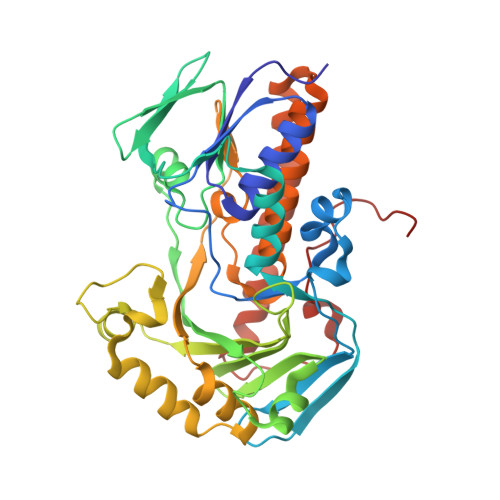
The enzyme 2,6-dihydroxypyridine-3-hydroxylase catalyzes the sixth step of the nicotine degradation pathway in Arthrobacter nicotinovorans. The enzyme was produced in Escherichia coli, purified and crystallized. The crystal structure was solved at 2.6 A resolution, revealing a significant structural relationship with the family of FAD-dependent aromatic hydroxylases, but essentially no sequence homology. The structure was aligned with those of the established family members, showing that the FAD molecules are bound at virtually identical locations. The reported enzyme is a dimer like most other family members, but its dimerization contact differs from the others. The binding position of NAD(P)H to this enzyme family is not clear. Since the reported enzyme accepts only NADH for flavin reduction in contrast to the other established members using NADPH, we searched through the structural alignment and found an indication for the position of the 2'-phosphate of NADPH that is in general agreement with mutational studies on a related enzyme, but contradicts a crystal soaking experiment. Using a bound glycerol molecule and the known substrate positions of three related enzymes as a guide, the substrate 2,6-dihydroxypyridine was placed into the active center. The access to the binding site is discussed. The new active center geometry introduces constraints that render some reaction scenarios more likely than others. It suggests that flavin is reduced at its out-position and then drawn into its in-position, where it binds molecular oxygen. The geometry is consistent with the proposal that peroxy-flavin is protonated by the solvent to yield the electrophilic hydroperoxy-flavin. The substrate is activated by two buried histidines but there is no appropriate base to store the surplus proton of the hydroxylated carbon atom. The implications of this problem are discussed.
Organizational Affiliation:
Institut für Organische Chemie und Biochemie, Albert-Ludwigs-Universität, Albertstr. 21, 79104 Freiburg im Breisgau, Germany.