2VFJ

Structure of the A20 Ovarian Tumour (OTU) domain


Experimental Data Snapshot

  • Method: X-RAY DIFFRACTION
  • Resolution: 3.20 Å
  • R-Value Free: 0.243 
  • R-Value Work: 0.204 
  • R-Value Observed: 0.206 

wwPDB Validation   3D Report Full Report


This is version 1.2 of the entry. See complete history


Literature

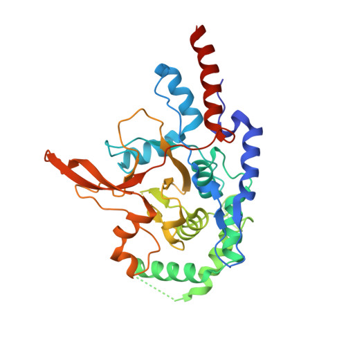
Structure of the A20 Otu Domain and Mechanistic Insights Into Deubiquitination

Komander, D.Barford, D.

(2008) Biochem J 409: 77

  • DOI: https://doi.org/10.1042/BJ20071399
  • Primary Citation of Related Structures:  
    2VFJ

  • PubMed Abstract: 

    The NF-kappaB (nuclear factor kappaB) regulator A20 antagonises IKK [IkappaB (inhibitor of kappaB) kinase] activation by modulating Lys63-linked polyubiquitination of cytokine-receptor-associated factors including TRAF2/6 (tumour-necrosis-factor-receptor-associated factor 2/6) and RIP1 (receptor-interacting protein 1). In the present paper we describe the crystal structure of the N-terminal OTU (ovarian tumour) deubiquitinase domain of A20, which differs from other deubiquitinases but shares the minimal catalytic core with otubain-2. Analysis of conserved surface regions allows prediction of ubiquitin-binding sites for the proximal and distal ubiquitin molecules. Structural and biochemical analysis suggests a novel architecture of the catalytic triad, which might be present in a subset of OTU domains including Cezanne and TRABID (TRAF-binding domain). Biochemical analysis shows a preference of the isolated A20 OTU domain for Lys48-linked tetraubiquitin in vitro suggesting that additional specificity factors might be required for the physiological function of A20 in cells.


  • Organizational Affiliation

    Section of Structural Biology, The Institute of Cancer Research, Chester Beatty Laboratories, 237 Fulham Road, London SW3 6JB, UK. david.komander@icr.ac.uk


Macromolecules
Find similar proteins by:  (by identity cutoff)  |  3D Structure
Entity ID: 1
MoleculeChains Sequence LengthOrganismDetailsImage
TUMOR NECROSIS FACTOR
A, B, C, D
366Homo sapiensMutation(s): 0 
EC: 3.4.19.12
UniProt & NIH Common Fund Data Resources
Find proteins for P21580 (Homo sapiens)
Explore P21580 
Go to UniProtKB:  P21580
PHAROS:  P21580
GTEx:  ENSG00000118503 
Entity Groups  
Sequence Clusters30% Identity50% Identity70% Identity90% Identity95% Identity100% Identity
UniProt GroupP21580
Sequence Annotations
Expand
  • Reference Sequence
Experimental Data & Validation

Experimental Data

  • Method: X-RAY DIFFRACTION
  • Resolution: 3.20 Å
  • R-Value Free: 0.243 
  • R-Value Work: 0.204 
  • R-Value Observed: 0.206 
  • Space Group: P 1 21 1
Unit Cell:
Length ( Å )Angle ( ˚ )
a = 84.964α = 90
b = 83.023β = 98.08
c = 164.94γ = 90
Software Package:
Software NamePurpose
PHENIXrefinement
MOSFLMdata reduction
SCALAdata scaling
SHELXCDEphasing
HKL2MAPphasing
SHARPphasing

Structure Validation

View Full Validation Report


Entry History 

Deposition Data

Revision History  (Full details and data files)

  • Version 1.0: 2007-12-04
    Type: Initial release
  • Version 1.1: 2011-05-08
    Changes: Version format compliance
  • Version 1.2: 2011-07-13
    Changes: Version format compliance