2VDI

Crystal structure of Chlamydomonas reinhardtii Rubisco with a large- subunit C192S mutation


Experimental Data Snapshot

  • Method: X-RAY DIFFRACTION
  • Resolution: 2.65 Å
  • R-Value Free: 0.234 
  • R-Value Work: 0.205 
  • R-Value Observed: 0.206 

wwPDB Validation   3D Report Full Report


Ligand Structure Quality Assessment 


This is version 1.4 of the entry. See complete history


Literature

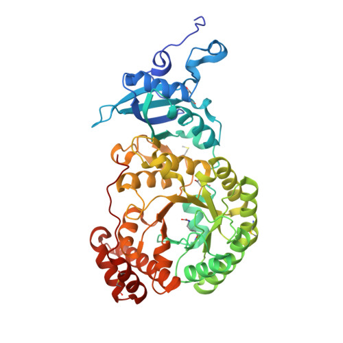
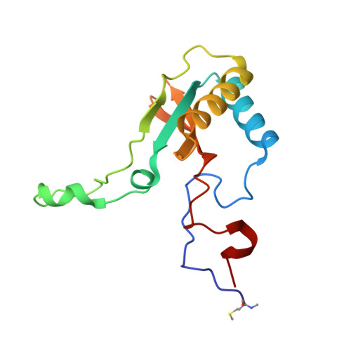
Structural and Functional Consequences of the Replacement of Proximal Residues Cys-172 and Cys-192 in the Large Subunit of Ribulose 1,5-Bisphosphate Carboxylase/Oxygenase from Chlamydomonas Reinhardtii

Garcia-Murria, M.-J.Karkehabadi, S.Marin-Navarro, J.Satagopan, S.Andersson, I.Spreitzer, R.J.Moreno, J.

(2008) Biochem J 411: 241

  • DOI: https://doi.org/10.1042/BJ20071422
  • Primary Citation of Related Structures:  
    2VDH, 2VDI

  • PubMed Abstract: 

    Proximal Cys(172) and Cys(192) in the large subunit of the photosynthetic enzyme Rubisco (ribulose-1,5-bisphosphate carboxylase/oxygenase; EC 4.1.1.39) are evolutionarily conserved among cyanobacteria, algae and higher plants. Mutation of Cys(172) has been shown to affect the redox properties of Rubisco in vitro and to delay the degradation of the enzyme in vivo under stress conditions. Here, we report the effect of the replacement of Cys(172) and Cys(192) by serine on the catalytic properties, thermostability and three-dimensional structure of Chlamydomonas reinhardtii Rubisco. The most striking effect of the C172S substitution was an 11% increase in the specificity factor when compared with the wild-type enzyme. The specificity factor of C192S Rubisco was not altered. The V(c) (V(max) for carboxylation) was similar to that of wild-type Rubisco in the case of the C172S enzyme, but approx. 30% lower for the C192S Rubisco. In contrast, the K(m) for CO(2) and O(2) was similar for C192S and wild-type enzymes, but distinctly higher (approximately double) for the C172S enzyme. C172S Rubisco showed a critical denaturation temperature approx. 2 degrees C lower than wild-type Rubisco and a distinctly higher denaturation rate at 55 degrees C, whereas C192S Rubisco was only slightly more sensitive to temperature denaturation than the wild-type enzyme. X-ray crystal structures reveal that the C172S mutation causes a shift of the main-chain backbone atoms of beta-strand 1 of the alpha/beta-barrel affecting a number of amino acid side chains. This may cause the exceptional catalytic features of C172S. In contrast, the C192S mutation does not produce similar structural perturbations.


  • Organizational Affiliation

    Departament de Bioquimica i Biologia Molecular, Facultat de Biologia, Universitat de València, Av. Dr Moliner 50, Burjassot, València E-46100, Spain.


Macromolecules
Find similar proteins by:  (by identity cutoff)  |  3D Structure
Entity ID: 1
MoleculeChains Sequence LengthOrganismDetailsImage
RIBULOSE BISPHOSPHATE CARBOXYLASE LARGE CHAIN
A, B, C, D, E
A, B, C, D, E, F, G, H
475Chlamydomonas reinhardtiiMutation(s): 1 
EC: 4.1.1.39
UniProt
Find proteins for P00877 (Chlamydomonas reinhardtii)
Explore P00877 
Go to UniProtKB:  P00877
Entity Groups  
Sequence Clusters30% Identity50% Identity70% Identity90% Identity95% Identity100% Identity
UniProt GroupP00877
Sequence Annotations
Expand
  • Reference Sequence
Find similar proteins by:  (by identity cutoff)  |  3D Structure
Entity ID: 2
MoleculeChains Sequence LengthOrganismDetailsImage
RIBULOSE BISPHOSPHATE CARBOXYLASE SMALL CHAIN 1
I, J, K, L, M
I, J, K, L, M, N, O, P
140Chlamydomonas reinhardtiiMutation(s): 0 
EC: 4.1.1.39
UniProt
Find proteins for P00873 (Chlamydomonas reinhardtii)
Explore P00873 
Go to UniProtKB:  P00873
Entity Groups  
Sequence Clusters30% Identity50% Identity70% Identity90% Identity95% Identity100% Identity
UniProt GroupP00873
Sequence Annotations
Expand
  • Reference Sequence
Small Molecules
Ligands 3 Unique
IDChains Name / Formula / InChI Key2D Diagram3D Interactions
CAP
Query on CAP

Download Ideal Coordinates CCD File 
DB [auth G]
EA [auth C]
JA [auth D]
KB [auth H]
OA [auth E]
DB [auth G],
EA [auth C],
JA [auth D],
KB [auth H],
OA [auth E],
R [auth A],
VA [auth F],
Y [auth B]
2-CARBOXYARABINITOL-1,5-DIPHOSPHATE
C6 H14 O13 P2
ITHCSGCUQDMYAI-ZMIZWQJLSA-N
EDO
Query on EDO

Download Ideal Coordinates CCD File 
AA [auth B]
AB [auth F]
BA [auth B]
BB [auth F]
CA [auth B]
AA [auth B],
AB [auth F],
BA [auth B],
BB [auth F],
CA [auth B],
EB [auth G],
FA [auth C],
FB [auth G],
GA [auth C],
GB [auth G],
HA [auth C],
HB [auth G],
IB [auth G],
KA [auth D],
LA [auth D],
LB [auth H],
MA [auth D],
MB [auth H],
NB [auth H],
OB [auth H],
PA [auth E],
PB [auth H],
QA [auth E],
QB [auth J],
RA [auth E],
RB [auth J],
S [auth A],
SA [auth E],
SB [auth K],
T [auth A],
TA [auth E],
TB [auth L],
U [auth A],
UB [auth N],
V [auth A],
VB [auth O],
W [auth A],
WA [auth F],
WB [auth O],
XA [auth F],
YA [auth F],
Z [auth B],
ZA [auth F]
1,2-ETHANEDIOL
C2 H6 O2
LYCAIKOWRPUZTN-UHFFFAOYSA-N
MG
Query on MG

Download Ideal Coordinates CCD File 
CB [auth G]
DA [auth C]
IA [auth D]
JB [auth H]
NA [auth E]
CB [auth G],
DA [auth C],
IA [auth D],
JB [auth H],
NA [auth E],
Q [auth A],
UA [auth F],
X [auth B]
MAGNESIUM ION
Mg
JLVVSXFLKOJNIY-UHFFFAOYSA-N
Modified Residues  4 Unique
IDChains TypeFormula2D DiagramParent
HYP
Query on HYP
A, B, C, D, E
A, B, C, D, E, F, G, H
L-PEPTIDE LINKINGC5 H9 N O3PRO
KCX
Query on KCX
A, B, C, D, E
A, B, C, D, E, F, G, H
L-PEPTIDE LINKINGC7 H14 N2 O4LYS
SMC
Query on SMC
A, B, C, D, E
A, B, C, D, E, F, G, H
L-PEPTIDE LINKINGC4 H9 N O2 SCYS
MME
Query on MME
I, J, K, L, M
I, J, K, L, M, N, O, P
L-PEPTIDE LINKINGC6 H13 N O2 SMET
Experimental Data & Validation

Experimental Data

  • Method: X-RAY DIFFRACTION
  • Resolution: 2.65 Å
  • R-Value Free: 0.234 
  • R-Value Work: 0.205 
  • R-Value Observed: 0.206 
  • Space Group: P 1 21 1
Unit Cell:
Length ( Å )Angle ( ˚ )
a = 120.831α = 90
b = 178.206β = 117.76
c = 122.917γ = 90
Software Package:
Software NamePurpose
REFMACrefinement
DENZOdata reduction
SCALEPACKdata scaling
AMoREphasing

Structure Validation

View Full Validation Report


Ligand Structure Quality Assessment 


Entry History 

Deposition Data

Revision History  (Full details and data files)

  • Version 1.0: 2008-11-04
    Type: Initial release
  • Version 1.1: 2011-05-08
    Changes: Version format compliance
  • Version 1.2: 2011-07-13
    Changes: Version format compliance
  • Version 1.3: 2018-01-17
    Changes: Data collection
  • Version 1.4: 2023-12-13
    Changes: Data collection, Database references, Derived calculations, Other, Refinement description