2VB3

Crystal structure of Ag(I)CusF


Experimental Data Snapshot

  • Method: X-RAY DIFFRACTION
  • Resolution: 2.33 Å
  • R-Value Free: 0.264 
  • R-Value Work: 0.241 
  • R-Value Observed: 0.242 

wwPDB Validation   3D Report Full Report


This is version 1.2 of the entry. See complete history


Literature

Cu(I) Recognition Via Cation-Pi and Methionine Interactions in Cusf.

Xue, Y.Davis, A.V.Balakrishnan, G.Stasser, J.P.Staehlin, B.M.Focia, P.Spiro, T.G.Penner-Hahn, J.E.O'Halloran, T.V.

(2008) Nat Chem Biol 4: 107

  • DOI: https://doi.org/10.1038/nchembio.2007.57
  • Primary Citation of Related Structures:  
    2VB2, 2VB3

  • PubMed Abstract: 

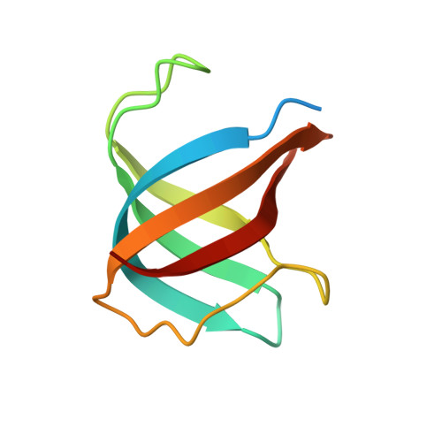
    Methionine-rich motifs have an important role in copper trafficking factors, including the CusF protein. Here we show that CusF uses a new metal recognition site wherein Cu(I) is tetragonally displaced from a Met2His ligand plane toward a conserved tryptophan. Spectroscopic studies demonstrate that both thioether ligation and strong cation-pi interactions with tryptophan stabilize metal binding. This novel active site chemistry affords mechanisms for control of adventitious metal redox and substitution chemistry.


  • Organizational Affiliation

    Department of Chemistry, Northwestern University, 2145 Sheridan Road, Evanston, Illinois 60208, USA.


Macromolecules
Find similar proteins by:  (by identity cutoff)  |  3D Structure
Entity ID: 1
MoleculeChains Sequence LengthOrganismDetailsImage
CATION EFFLUX SYSTEM PROTEIN CUSFA [auth X]88Escherichia coliMutation(s): 0 
UniProt
Find proteins for P77214 (Escherichia coli (strain K12))
Explore P77214 
Go to UniProtKB:  P77214
Entity Groups  
Sequence Clusters30% Identity50% Identity70% Identity90% Identity95% Identity100% Identity
UniProt GroupP77214
Sequence Annotations
Expand
  • Reference Sequence
Small Molecules
Ligands 1 Unique
IDChains Name / Formula / InChI Key2D Diagram3D Interactions
AG
Query on AG

Download Ideal Coordinates CCD File 
B [auth X]SILVER ION
Ag
FOIXSVOLVBLSDH-UHFFFAOYSA-N
Experimental Data & Validation

Experimental Data

  • Method: X-RAY DIFFRACTION
  • Resolution: 2.33 Å
  • R-Value Free: 0.264 
  • R-Value Work: 0.241 
  • R-Value Observed: 0.242 
  • Space Group: P 21 21 21
Unit Cell:
Length ( Å )Angle ( ˚ )
a = 39.658α = 90
b = 44.229β = 90
c = 44.241γ = 90
Software Package:
Software NamePurpose
REFMACrefinement
MOSFLMdata reduction
SCALAdata scaling
PHASERphasing

Structure Validation

View Full Validation Report


Entry History 

Deposition Data

Revision History  (Full details and data files)

  • Version 1.0: 2007-12-18
    Type: Initial release
  • Version 1.1: 2011-07-13
    Changes: Advisory, Version format compliance
  • Version 1.2: 2023-12-13
    Changes: Data collection, Database references, Refinement description