2V76

Crystal structure of the human dok1 PTB domain


Experimental Data Snapshot

  • Method: X-RAY DIFFRACTION
  • Resolution: 1.60 Å
  • R-Value Free: 0.224 
  • R-Value Work: 0.188 
  • R-Value Observed: 0.190 

wwPDB Validation   3D Report Full Report


This is version 1.3 of the entry. See complete history


Literature

An Integrin Phosphorylation Switch: The Effect of {Beta}3 Integrin Tail Phosphorylation on Dok1 and Talin Binding.

Oxley, C.L.Anthis, N.J.Lowe, E.D.Vakonakis, I.Campbell, I.D.Wegener, K.L.

(2008) J Biol Chem 283: 5420

  • DOI: https://doi.org/10.1074/jbc.M709435200
  • Primary Citation of Related Structures:  
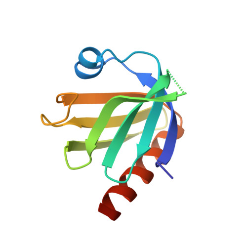
    2V76

  • PubMed Abstract: 

    Integrins play a fundamental role in cell migration and adhesion; knowledge of how they are regulated and controlled is vital for understanding these processes. Recent work showed that Dok1 negatively regulates integrin activation, presumably by competition with talin. To understand how this occurs, we used NMR spectroscopy and x-ray crystallography to investigate the molecular details of interactions with integrins. The binding affinities of beta3 integrin tails for the Dok1 and talin phosphotyrosine binding domains were quantified using 15N-1H hetero-nuclear single quantum correlation titrations, revealing that the unphosphorylated integrin tail binds more strongly to talin than Dok1. Chemical shift mapping showed that unlike talin, Dok1 exclusively interacts with the canonical NPXY motif of the beta3 integrin tail. Upon phosphorylation of Tyr 747 in the beta3 integrin tail, however, Dok1 then binds much more strongly than talin. Thus, we show that phosphorylation of Tyr 747 provides a switch for integrin ligand binding. This switch may represent an in vivo mechanism for control of integrin receptor activation. These results have implications for the control of integrin signaling by proteins containing phosphotyrosine binding domains.


  • Organizational Affiliation

    Department of Biochemistry, University of Oxford, South Parks Road, Oxford OX1 3QU, United Kingdom.


Macromolecules
Find similar proteins by:  (by identity cutoff)  |  3D Structure
Entity ID: 1
MoleculeChains Sequence LengthOrganismDetailsImage
DOCKING PROTEIN 1
A, B, C, D
107Homo sapiensMutation(s): 0 
UniProt & NIH Common Fund Data Resources
Find proteins for Q99704 (Homo sapiens)
Explore Q99704 
Go to UniProtKB:  Q99704
PHAROS:  Q99704
GTEx:  ENSG00000115325 
Entity Groups  
Sequence Clusters30% Identity50% Identity70% Identity90% Identity95% Identity100% Identity
UniProt GroupQ99704
Sequence Annotations
Expand
  • Reference Sequence
Small Molecules
Ligands 4 Unique
IDChains Name / Formula / InChI Key2D Diagram3D Interactions
PGE
Query on PGE

Download Ideal Coordinates CCD File 
E [auth A],
H [auth B],
I [auth B],
M [auth D]
TRIETHYLENE GLYCOL
C6 H14 O4
ZIBGPFATKBEMQZ-UHFFFAOYSA-N
SO4
Query on SO4

Download Ideal Coordinates CCD File 
F [auth A],
J [auth B],
K [auth C],
N [auth D]
SULFATE ION
O4 S
QAOWNCQODCNURD-UHFFFAOYSA-L
GOL
Query on GOL

Download Ideal Coordinates CCD File 
L [auth C]GLYCEROL
C3 H8 O3
PEDCQBHIVMGVHV-UHFFFAOYSA-N
EDO
Query on EDO

Download Ideal Coordinates CCD File 
G [auth A]1,2-ETHANEDIOL
C2 H6 O2
LYCAIKOWRPUZTN-UHFFFAOYSA-N
Experimental Data & Validation

Experimental Data

  • Method: X-RAY DIFFRACTION
  • Resolution: 1.60 Å
  • R-Value Free: 0.224 
  • R-Value Work: 0.188 
  • R-Value Observed: 0.190 
  • Space Group: C 1 2 1
Unit Cell:
Length ( Å )Angle ( ˚ )
a = 76.88α = 90
b = 100.168β = 117.34
c = 67.101γ = 90
Software Package:
Software NamePurpose
REFMACrefinement
MOSFLMdata reduction
SCALAdata scaling
PHASERphasing

Structure Validation

View Full Validation Report


Entry History 

Deposition Data

Revision History  (Full details and data files)

  • Version 1.0: 2008-01-08
    Type: Initial release
  • Version 1.1: 2011-05-08
    Changes: Version format compliance
  • Version 1.2: 2011-07-13
    Changes: Version format compliance
  • Version 1.3: 2023-12-13
    Changes: Data collection, Database references, Other, Refinement description