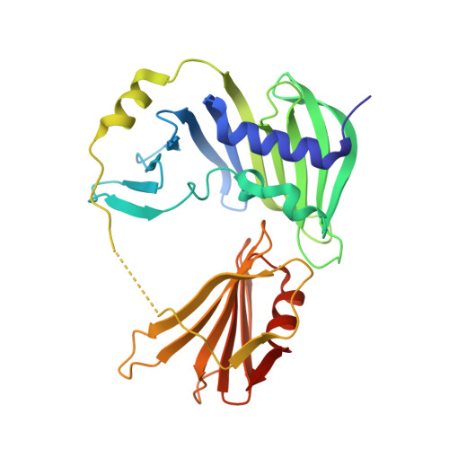
2V42

Crystal structure of RseB: a sensor for periplasmic stress response in E. coli


Experimental Data Snapshot

  • Method: X-RAY DIFFRACTION
  • Resolution: 2.75 Å
  • R-Value Free: 0.258 
  • R-Value Work: 0.217 
  • R-Value Observed: 0.219 

wwPDB Validation   3D Report Full Report


Ligand Structure Quality Assessment 


This is version 1.1 of the entry. See complete history


Literature

The Structure of Rseb: A Sensor in Periplasmic Stress Response of E. Coli.

Wollmann, P.Zeth, K.

(2007) J Mol Biol 372: 927

  • DOI: https://doi.org/10.1016/j.jmb.2007.06.039
  • Primary Citation of Related Structures:  
    2V42, 2V43

  • PubMed Abstract: 

    An elegant network of signal transduction has evolved in the bacterial cell envelope to respond to environmental stress. It is initiated by sensing unfavourable and harmful changes in the periplasm. The stress signal is then transmitted by a controlled degradation of the transmembrane anti-sigma-factor RseA that leads to the activation of the alternative sigma factor sigma(E). The periplasmic protein RseB exerts a crucial role in modulating the stability of RseA. RseB from Escherichia coli has been crystallized and crystal structures were determined at 2.4 A and at 2.8 A resolution. The protein forms a homodimer, with the monomer composed of two domains. The large domain resembles an unclosed beta-barrel that is structurally remarkably similar to a protein family capable of binding the lipid anchor of lipoproteins. The small C-terminal domain, connected to the large domain by a partially unstructured loop, is responsible for interaction with RseA. On the basis of the structure of RseB, we suggest that it acts as a sensor of periplasmic stress with a dual functionality: it detects mislocalized lipoproteins and propagates the signal to induce the sigma(E)-response.


  • Organizational Affiliation

    Max Planck Institute of Biochemistry, Department of Membrane Biochemistry, Am Klopferspitz 18, D-82152 Martinsried, Germany.


Macromolecules
Find similar proteins by:  (by identity cutoff)  |  3D Structure
Entity ID: 1
MoleculeChains Sequence LengthOrganismDetailsImage
SIGMA-E FACTOR REGULATORY PROTEIN RSEB
A, B
303Escherichia coli BL21(DE3)Mutation(s): 0 
UniProt
Find proteins for P0AFX9 (Escherichia coli (strain K12))
Explore P0AFX9 
Go to UniProtKB:  P0AFX9
Entity Groups  
Sequence Clusters30% Identity50% Identity70% Identity90% Identity95% Identity100% Identity
UniProt GroupP0AFX9
Sequence Annotations
Expand
  • Reference Sequence
Small Molecules
Ligands 1 Unique
IDChains Name / Formula / InChI Key2D Diagram3D Interactions
NDS
Query on NDS

Download Ideal Coordinates CCD File 
C [auth A],
D [auth B]
ETHYL DIMETHYL AMMONIO PROPANE SULFONATE
C7 H17 N O3 S
NNCRHRDBFDCWPA-UHFFFAOYSA-N
Experimental Data & Validation

Experimental Data

  • Method: X-RAY DIFFRACTION
  • Resolution: 2.75 Å
  • R-Value Free: 0.258 
  • R-Value Work: 0.217 
  • R-Value Observed: 0.219 
  • Space Group: P 4 21 2
Unit Cell:
Length ( Å )Angle ( ˚ )
a = 164.29α = 90
b = 164.29β = 90
c = 81.52γ = 90
Software Package:
Software NamePurpose
REFMACrefinement
XDSdata reduction
XSCALEdata scaling

Structure Validation

View Full Validation Report


Ligand Structure Quality Assessment 


Entry History 

Deposition Data

Revision History  (Full details and data files)

  • Version 1.0: 2007-08-28
    Type: Initial release
  • Version 1.1: 2011-07-13
    Changes: Advisory, Version format compliance