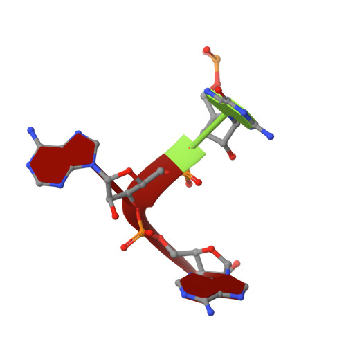
2TMV

VISUALIZATION OF PROTEIN-NUCLEIC ACID INTERACTIONS IN A VIRUS. REFINED STRUCTURE OF INTACT TOBACCO MOSAIC VIRUS AT 2.9 ANGSTROMS RESOLUTION BY X-RAY FIBER DIFFRACTION


Experimental Data Snapshot

  • Method: FIBER DIFFRACTION
  • Resolution: 2.90 Å
  • R-Value Observed: 0.096 

wwPDB Validation   3D Report Full Report


This is version 1.4 of the entry. See complete history


Literature

Visualization of protein-nucleic acid interactions in a virus. Refined structure of intact tobacco mosaic virus at 2.9 A resolution by X-ray fiber diffraction.

Namba, K.Pattanayek, R.Stubbs, G.

(1989) J Mol Biol 208: 307-325

  • DOI: https://doi.org/10.1016/0022-2836(89)90391-4
  • Primary Citation of Related Structures:  
    2TMV

  • PubMed Abstract: 

    The structure of tobacco mosaic virus (TMV) has been determined by fiber diffraction methods at 2.9 A resolution, and refined by restrained least-squares to an R-factor of 0.096. Protein-nucleic acid interactions are clearly visible. The final model contains all of the non-hydrogen atoms of the RNA and the protein, 71 water molecules, and two calcium-binding sites. Viral disassembly is driven by electrostatic repulsions between the charges in two carboxyl-carboxylate pairs and a phosphate-carboxylate pair. The phosphate-carboxylate pair and at least one of the carboxyl-carboxylate pairs appear to be calcium-binding sites. Nucleotide specificity, enabling TMV to recognize its own RNA by a repeating pattern of guanine residues, is provided by two guanine-specific hydrogen bonds in one of the three base-binding sites.


  • Organizational Affiliation

    Department of Molecular Biology, Vanderbilt University, Nashville, TN 37235.


Macromolecules

Find similar proteins by:  (by identity cutoff)  |  3D Structure
Entity ID: 2
MoleculeChains Sequence LengthOrganismDetailsImage
TMV COAT PROTEINB [auth P]158Tobacco mosaic virus (vulgare)Mutation(s): 0 
UniProt
Find proteins for P69687 (Tobacco mosaic virus (strain vulgare))
Explore P69687 
Go to UniProtKB:  P69687
Entity Groups  
Sequence Clusters30% Identity50% Identity70% Identity90% Identity95% Identity100% Identity
UniProt GroupP69687
Sequence Annotations
Expand
  • Reference Sequence

Find similar nucleic acids by:  Sequence   |   3D Structure  

Entity ID: 1
MoleculeChains LengthOrganismImage
RNA (5'-R(P*GP*AP*A)-3')A [auth R]3N/A
Sequence Annotations
Expand
  • Reference Sequence
Small Molecules
Ligands 1 Unique
IDChains Name / Formula / InChI Key2D Diagram3D Interactions
CA
Query on CA

Download Ideal Coordinates CCD File 
C [auth R]CALCIUM ION
Ca
BHPQYMZQTOCNFJ-UHFFFAOYSA-N
Experimental Data & Validation

Experimental Data

  • Method: FIBER DIFFRACTION
  • Resolution: 2.90 Å
  • R-Value Observed: 0.096 
  • Space Group: P 1
Unit Cell:
Length ( Å )Angle ( ˚ )
a = 1α = 90
b = 1β = 90
c = 1γ = 90
Software Package:
Software NamePurpose
PROLSQrefinement

Structure Validation

View Full Validation Report


Entry History 

Deposition Data

Revision History  (Full details and data files)

  • Version 1.0: 1989-01-09
    Type: Initial release
  • Version 1.1: 2008-03-25
    Changes: Version format compliance
  • Version 1.2: 2011-07-13
    Changes: Version format compliance
  • Version 1.3: 2017-11-29
    Changes: Derived calculations, Other
  • Version 1.4: 2024-02-21
    Changes: Data collection, Database references, Derived calculations