2RIA

Crystal structure of the trimeric neck and carbohydrate recognition domain of human surfactant protein D in complex with D-glycero-D-manno-heptose


Experimental Data Snapshot

  • Method: X-RAY DIFFRACTION
  • Resolution: 1.80 Å
  • R-Value Free: 0.237 
  • R-Value Work: 0.209 

wwPDB Validation   3D Report Full Report


Ligand Structure Quality Assessment 


This is version 1.3 of the entry. See complete history


Literature

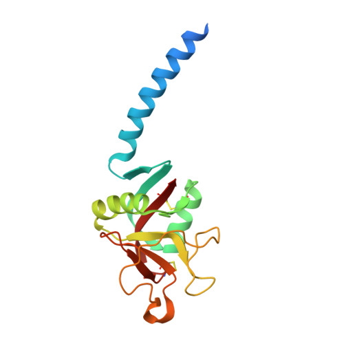
Recognition of heptoses and the inner core of bacterial lipopolysaccharides by surfactant protein d.

Wang, H.Head, J.Kosma, P.Brade, H.Muller-Loennies, S.Sheikh, S.McDonald, B.Smith, K.Cafarella, T.Seaton, B.Crouch, E.

(2008) Biochemistry 47: 710-720

  • DOI: https://doi.org/10.1021/bi7020553
  • Primary Citation of Related Structures:  
    2RIA, 2RIB, 2RIC, 2RID, 2RIE

  • PubMed Abstract: 

    Lipopolysaccharides (LPS) of Gram-negative bacteria are important mediators of bacterial virulence that can elicit potent endotoxic effects. Surfactant protein D (SP-D) shows specific interactions with LPS, both in vitro and in vivo. These interactions involve binding of the carbohydrate recognition domain (CRD) to LPS oligosaccharides (OS); however, little is known about the mechanisms of LPS recognition. Recombinant neck+CRDs (NCRDs) provide an opportunity to directly correlate binding interactions with a crystallographic analysis of the binding mechanism. In these studies, we examined the interactions of wild-type and mutant trimeric NCRDs with rough LPS (R-LPS). Although rat NCRDs bound more efficiently than human NCRDs to Escherichia coli J-5 LPS, both proteins exhibited efficient binding to solid-phase Rd2-LPS and to Rd2-LPS aggregates presented in the solution phase. Involvement of residues flanking calcium at the sugar binding site was demonstrated by reciprocal exchange of lysine and arginine at position 343 of rat and human CRDs. The lectin activity of hNCRDs was inhibited by specific heptoses, including l-glycero-alpha-d-manno-heptose (l,d-heptose), but not by 3-deoxy-alpha-d-manno-oct-2-ulosonic acid (Kdo). Crystallographic analysis of the hNCRD demonstrated a novel binding orientation for l,d-heptose, involving the hydroxyl groups of the side chain. Similar binding was observed for a synthetic alpha1-->3-linked heptose disaccharide corresponding to heptoses I and II of the inner core region in many LPS. 7-O-Carbamoyl-l,d-heptose and d-glycero-alpha-d-manno-heptose were bound via ring hydroxyl groups. Interactions with the side chain of inner core heptoses provide a potential mechanism for the recognition of diverse types of LPS by SP-D.


  • Organizational Affiliation

    Department Physiology and Biophysics, Boston University School of Medicine, Boston, Massachusetts 02118, USA.


Macromolecules
Find similar proteins by:  (by identity cutoff)  |  3D Structure
Entity ID: 1
MoleculeChains Sequence LengthOrganismDetailsImage
Pulmonary surfactant-associated protein D
A, B, C
160Homo sapiensMutation(s): 0 
Gene Names: SFTPDPSPDSFTP4
UniProt & NIH Common Fund Data Resources
Find proteins for P35247 (Homo sapiens)
Explore P35247 
Go to UniProtKB:  P35247
PHAROS:  P35247
GTEx:  ENSG00000133661 
Entity Groups  
Sequence Clusters30% Identity50% Identity70% Identity90% Identity95% Identity100% Identity
UniProt GroupP35247
Sequence Annotations
Expand
  • Reference Sequence
Small Molecules
Ligands 2 Unique
IDChains Name / Formula / InChI Key2D Diagram3D Interactions
289
Query on 289

Download Ideal Coordinates CCD File 
G [auth A],
K [auth B],
O [auth C]
D-glycero-alpha-D-manno-heptopyranose
C7 H14 O7
BGWQRWREUZVRGI-QTNLNCNHSA-N
CA
Query on CA

Download Ideal Coordinates CCD File 
D [auth A]
E [auth A]
F [auth A]
H [auth B]
I [auth B]
D [auth A],
E [auth A],
F [auth A],
H [auth B],
I [auth B],
J [auth B],
L [auth C],
M [auth C],
N [auth C]
CALCIUM ION
Ca
BHPQYMZQTOCNFJ-UHFFFAOYSA-N
Binding Affinity Annotations 
IDSourceBinding Affinity
289 PDBBind:  2RIA IC50: 2.70e+6 (nM) from 1 assay(s)
Binding MOAD:  2RIA IC50: 2.70e+6 (nM) from 1 assay(s)
Experimental Data & Validation

Experimental Data

  • Method: X-RAY DIFFRACTION
  • Resolution: 1.80 Å
  • R-Value Free: 0.237 
  • R-Value Work: 0.209 
  • Space Group: P 1 21 1
Unit Cell:
Length ( Å )Angle ( ˚ )
a = 55.414α = 90
b = 108.606β = 91.03
c = 55.548γ = 90
Software Package:
Software NamePurpose
DENZOdata reduction
SCALEPACKdata scaling
EPMRphasing
CNSrefinement
PDB_EXTRACTdata extraction
CrystalCleardata collection
CNSphasing

Structure Validation

View Full Validation Report


Ligand Structure Quality Assessment 


Entry History 

Deposition Data

Revision History  (Full details and data files)

  • Version 1.0: 2008-01-15
    Type: Initial release
  • Version 1.1: 2011-07-13
    Changes: Version format compliance
  • Version 1.2: 2017-10-25
    Changes: Refinement description
  • Version 1.3: 2020-07-29
    Type: Remediation
    Reason: Carbohydrate remediation
    Changes: Data collection, Database references, Derived calculations