2REB

THE STRUCTURE OF THE E. COLI RECA PROTEIN MONOMER AND POLYMER


Experimental Data Snapshot

  • Method: X-RAY DIFFRACTION
  • Resolution: 2.30 Å
  • R-Value Work: 0.210 
  • R-Value Observed: 0.210 

wwPDB Validation   3D Report Full Report


This is version 1.4 of the entry. See complete history


Literature

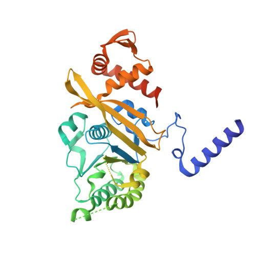
The structure of the E. coli recA protein monomer and polymer.

Story, R.M.Weber, I.T.Steitz, T.A.

(1992) Nature 355: 318-325

  • DOI: https://doi.org/10.1038/355318a0
  • Primary Citation of Related Structures:  
    2REB

  • PubMed Abstract: 

    The crystal structure of the recA protein from Escherichia coli at 2.3-A resolution reveals a major domain that binds ADP and probably single- and double-stranded DNA. Two smaller subdomains at the N and C termini protrude from the protein and respectively stabilize a 6(1) helical polymer of protein subunits and interpolymer bundles. This polymer structure closely resembles that of recA/DNA filaments determined by electron microscopy. Mutations in recA protein that enhance coprotease, DNA-binding and/or strand-exchange activity can be explained if the interpolymer interactions in the crystal reflect a regulatory mechanism in vivo.


  • Organizational Affiliation

    Department of Molecular Biophysics and Biochemistry, Yale University, New Haven, Connecticut 06511.


Macromolecules
Find similar proteins by:  (by identity cutoff)  |  3D Structure
Entity ID: 1
MoleculeChains Sequence LengthOrganismDetailsImage
REC A352Escherichia coliMutation(s): 0 
UniProt
Find proteins for P0A7G6 (Escherichia coli (strain K12))
Explore P0A7G6 
Go to UniProtKB:  P0A7G6
Entity Groups  
Sequence Clusters30% Identity50% Identity70% Identity90% Identity95% Identity100% Identity
UniProt GroupP0A7G6
Sequence Annotations
Expand
  • Reference Sequence
Experimental Data & Validation

Experimental Data

  • Method: X-RAY DIFFRACTION
  • Resolution: 2.30 Å
  • R-Value Work: 0.210 
  • R-Value Observed: 0.210 
  • Space Group: P 61
Unit Cell:
Length ( Å )Angle ( ˚ )
a = 102.4α = 90
b = 102.4β = 90
c = 82.7γ = 120
Software Package:
Software NamePurpose
X-PLORmodel building
X-PLORrefinement
X-PLORphasing

Structure Validation

View Full Validation Report


Entry History 

Deposition Data

Revision History  (Full details and data files)

  • Version 1.0: 1993-10-31
    Type: Initial release
  • Version 1.1: 2008-03-25
    Changes: Version format compliance
  • Version 1.2: 2011-07-13
    Changes: Version format compliance
  • Version 1.3: 2012-05-09
    Changes: Other
  • Version 1.4: 2024-02-21
    Changes: Data collection, Database references, Other