2RE3

CRYSTAL STRUCTURE OF a DUF1285 family protein (SPO_0140) FROM SILICIBACTER POMEROYI DSS-3 AT 2.50 A RESOLUTION


Experimental Data Snapshot

  • Method: X-RAY DIFFRACTION
  • Resolution: 2.50 Å
  • R-Value Free: 0.258 
  • R-Value Work: 0.215 
  • R-Value Observed: 0.217 

wwPDB Validation   3D Report Full Report


This is version 1.4 of the entry. See complete history


Literature

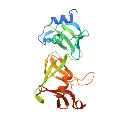
Structures of the first representatives of Pfam family PF06938 (DUF1285) reveal a new fold with repeated structural motifs and possible involvement in signal transduction.

Han, G.W.Bakolitsa, C.Miller, M.D.Kumar, A.Carlton, D.Najmanovich, R.J.Abdubek, P.Astakhova, T.Axelrod, H.L.Chen, C.Chiu, H.J.Clayton, T.Das, D.Deller, M.C.Duan, L.Ernst, D.Feuerhelm, J.Grant, J.C.Grzechnik, A.Jaroszewski, L.Jin, K.K.Johnson, H.A.Klock, H.E.Knuth, M.W.Kozbial, P.Krishna, S.S.Marciano, D.McMullan, D.Morse, A.T.Nigoghossian, E.Okach, L.Reyes, R.Rife, C.L.Sefcovic, N.Tien, H.J.Trame, C.B.van den Bedem, H.Weekes, D.Xu, Q.Hodgson, K.O.Wooley, J.Elsliger, M.A.Deacon, A.M.Godzik, A.Lesley, S.A.Wilson, I.A.

(2010) Acta Crystallogr Sect F Struct Biol Cryst Commun 66: 1218-1225

  • DOI: https://doi.org/10.1107/S1744309109050416
  • Primary Citation of Related Structures:  
    2RA9, 2RE3

  • PubMed Abstract: 

    The crystal structures of SPO0140 and Sbal_2486 were determined using the semiautomated high-throughput pipeline of the Joint Center for Structural Genomics (JCSG) as part of the NIGMS Protein Structure Initiative (PSI). The structures revealed a conserved core with domain duplication and a superficial similarity of the C-terminal domain to pleckstrin homology-like folds. The conservation of the domain interface indicates a potential binding site that is likely to involve a nucleotide-based ligand, with genome-context and gene-fusion analyses additionally supporting a role for this family in signal transduction, possibly during oxidative stress.


  • Organizational Affiliation

    Stanford Synchrotron Radiation ightsource, SLAC National Accelerator Laboratory, Menlo Park, CA, USA.


Macromolecules
Find similar proteins by:  (by identity cutoff)  |  3D Structure
Entity ID: 1
MoleculeChains Sequence LengthOrganismDetailsImage
Uncharacterized protein
A, B
194Ruegeria pomeroyi DSS-3Mutation(s): 0 
Gene Names: YP_165412.1SPO0140
UniProt
Find proteins for Q5LWU5 (Ruegeria pomeroyi (strain ATCC 700808 / DSM 15171 / DSS-3))
Explore Q5LWU5 
Go to UniProtKB:  Q5LWU5
Entity Groups  
Sequence Clusters30% Identity50% Identity70% Identity90% Identity95% Identity100% Identity
UniProt GroupQ5LWU5
Sequence Annotations
Expand
  • Reference Sequence
Small Molecules
Ligands 1 Unique
IDChains Name / Formula / InChI Key2D Diagram3D Interactions
GOL
Query on GOL

Download Ideal Coordinates CCD File 
C [auth A]GLYCEROL
C3 H8 O3
PEDCQBHIVMGVHV-UHFFFAOYSA-N
Modified Residues  1 Unique
IDChains TypeFormula2D DiagramParent
MSE
Query on MSE
A, B
L-PEPTIDE LINKINGC5 H11 N O2 SeMET
Experimental Data & Validation

Experimental Data

Unit Cell:
Length ( Å )Angle ( ˚ )
a = 75.37α = 90
b = 75.37β = 90
c = 182.687γ = 90
Software Package:
Software NamePurpose
REFMACrefinement
PHENIXrefinement
SHARPphasing
MolProbitymodel building
SCALAdata scaling
PDB_EXTRACTdata extraction
MAR345data collection
MOSFLMdata reduction

Structure Validation

View Full Validation Report


Entry History 

Deposition Data

Revision History  (Full details and data files)

  • Version 1.0: 2007-10-09
    Type: Initial release
  • Version 1.1: 2011-07-13
    Changes: Advisory, Version format compliance
  • Version 1.2: 2017-10-25
    Changes: Author supporting evidence, Refinement description
  • Version 1.3: 2019-07-24
    Changes: Data collection, Derived calculations, Refinement description
  • Version 1.4: 2023-01-25
    Changes: Database references, Derived calculations