Active Site and Loop 4 Movements within Human Glycolate Oxidase: Implications for Substrate Specificity and Drug Design.
Murray, M.S., Holmes, R.P., Lowther, W.T.(2008) Biochemistry 47: 2439-2449
- PubMed: 18215067
- DOI: https://doi.org/10.1021/bi701710r
- Primary Citation of Related Structures:
2RDT, 2RDU, 2RDW - PubMed Abstract:
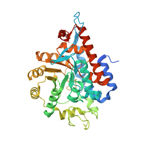
Human glycolate oxidase (GO) catalyzes the FMN-dependent oxidation of glycolate to glyoxylate and glyoxylate to oxalate, a key metabolite in kidney stone formation. We report herein the structures of recombinant GO complexed with sulfate, glyoxylate, and an inhibitor, 4-carboxy-5-dodecylsulfanyl-1,2,3-triazole (CDST), determined by X-ray crystallography. In contrast to most alpha-hydroxy acid oxidases including spinach glycolate oxidase, a loop region, known as loop 4, is completely visible when the GO active site contains a small ligand. The lack of electron density for this loop in the GO-CDST complex, which mimics a large substrate, suggests that a disordered to ordered transition may occur with the binding of substrates. The conformational flexibility of Trp110 appears to be responsible for enabling GO to react with alpha-hydroxy acids of various chain lengths. Moreover, the movement of Trp110 disrupts a hydrogen-bonding network between Trp110, Leu191, Tyr134, and Tyr208. This loss of interactions is the first indication that active site movements are directly linked to changes in the conformation of loop 4. The kinetic parameters for the oxidation of glycolate, glyoxylate, and 2-hydroxy octanoate indicate that the oxidation of glycolate to glyoxylate is the primary reaction catalyzed by GO, while the oxidation of glyoxylate to oxalate is most likely not relevant under normal conditions. However, drugs that exploit the unique structural features of GO may ultimately prove to be useful for decreasing glycolate and glyoxylate levels in primary hyperoxaluria type 1 patients who have the inability to convert peroxisomal glyoxylate to glycine.
Organizational Affiliation:
Center for Structural Biology and Department of Biochemistry, Wake Forest University Health Sciences, Medical Center Boulevard, Winston-Salem, North Carolina 27157, USA.