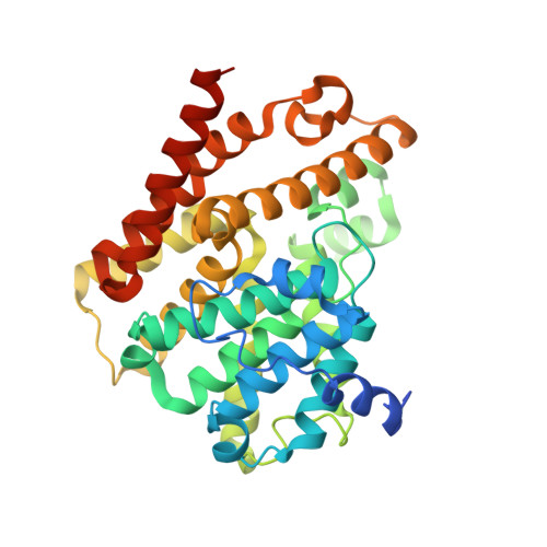
Crystal structure of the Leishmania major phosphodiesterase LmjPDEB1 and insight into the design of the parasite-selective inhibitors.
Wang, H., Yan, Z., Geng, J., Kunz, S., Seebeck, T., Ke, H.(2007) Mol Microbiol 66: 1029-1038
- PubMed: 17944832
- DOI: https://doi.org/10.1111/j.1365-2958.2007.05976.x
- Primary Citation of Related Structures:
2R8Q - PubMed Abstract:
Human leishmaniasis is a major public health problem in many countries, but chemotherapy is in an unsatisfactory state. Leishmania major phosphodiesterases (LmjPDEs) have been shown to play important roles in cell proliferation and apoptosis of the parasite. Thus LmjPDE inhibitors may potentially represent a novel class of drugs for the treatment of leishmaniasis. Reported here are the kinetic characterization of the LmjPDEB1 catalytic domain and its crystal structure as a complex with 3-isobutyl-1-methylxanthine (IBMX) at 1.55 A resolution. The structure of LmjPDEB1 is similar to that of human PDEs. IBMX stacks against the conserved phenylalanine and forms a hydrogen bond with the invariant glutamine, in a pattern common to most inhibitors bound to human PDEs. However, an extensive structural comparison reveals subtle, but significant differences between the active sites of LmjPDEB1 and human PDEs. In addition, a pocket next to the inhibitor binding site is found to be unique to LmjPDEB1. This pocket is isolated by two gating residues in human PDE families, but constitutes a natural expansion of the inhibitor binding pocket in LmjPDEB1. The structure particularity might be useful for the development of parasite-selective inhibitors for the treatment of leishmaniasis.
Organizational Affiliation:
Department of Biochemistry and Biophysics and Lineberger Comprehensive Cancer Center, The University of North Carolina, Chapel Hill, NC 27599-7260, USA.