2R4X

Ligand Migration and Binding in The Dimeric Hemoglobin of Scapharca Inaequivalvis: H69V/I114M co complex


Experimental Data Snapshot

  • Method: X-RAY DIFFRACTION
  • Resolution: 2.10 Å
  • R-Value Free: 0.210 
  • R-Value Work: 0.186 
  • R-Value Observed: 0.186 

wwPDB Validation   3D Report Full Report


Ligand Structure Quality Assessment 


This is version 1.4 of the entry. See complete history


Literature

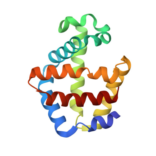
Ligand Migration and Binding in the Dimeric Hemoglobin of Scapharca inaequivalvis

Nienhaus, K.Knapp, J.E.Palladino, P.Royer Jr., W.E.Nienhaus, G.U.

(2007) Biochemistry 46: 14018-14031

  • DOI: https://doi.org/10.1021/bi7016798
  • Primary Citation of Related Structures:  
    2R4W, 2R4X, 2R4Y, 2R4Z, 2Z85, 2Z8A

  • PubMed Abstract: 

    Using Fourier transform infrared (FTIR) spectroscopy combined with temperature derivative spectroscopy (TDS) at cryogenic temperatures, we have studied CO binding to the heme and CO migration among cavities in the interior of the dimeric hemoglobin of Scapharca inaequivalvis (HbI) after photodissociation. By combining these studies with X-ray crystallography, three transient ligand docking sites were identified: a primary docking site B in close vicinity to the heme iron, and two secondary docking sites C and D corresponding to the Xe4 and Xe2 cavities of myoglobin. To assess the relevance of these findings for physiological binding, we also performed flash photolysis experiments on HbICO at room temperature and equilibrium binding studies with dioxygen. Our results show that the Xe4 and Xe2 cavities serve as transient docking sites for unbound ligands in the protein, but not as way stations on the entry/exit pathway. For HbI, the so-called histidine gate mechanism proposed for other globins appears as a plausible entry/exit route as well.


  • Organizational Affiliation

    Institute of Biophysics, University of Ulm, 89069 Ulm, Germany.


Macromolecules
Find similar proteins by:  (by identity cutoff)  |  3D Structure
Entity ID: 1
MoleculeChains Sequence LengthOrganismDetailsImage
Globin-1
A, B
146Anadara inaequivalvisMutation(s): 2 
Gene Names: HbI
UniProt
Find proteins for P02213 (Anadara inaequivalvis)
Explore P02213 
Go to UniProtKB:  P02213
Entity Groups  
Sequence Clusters30% Identity50% Identity70% Identity90% Identity95% Identity100% Identity
UniProt GroupP02213
Sequence Annotations
Expand
  • Reference Sequence
Experimental Data & Validation

Experimental Data

  • Method: X-RAY DIFFRACTION
  • Resolution: 2.10 Å
  • R-Value Free: 0.210 
  • R-Value Work: 0.186 
  • R-Value Observed: 0.186 
  • Space Group: C 2 2 21
Unit Cell:
Length ( Å )Angle ( ˚ )
a = 92.25α = 90
b = 44.37β = 90
c = 143.86γ = 90
Software Package:
Software NamePurpose
CNSrefinement
MAR345data collection
DENZOdata reduction
SCALEPACKdata scaling
CNSphasing

Structure Validation

View Full Validation Report


Ligand Structure Quality Assessment 


Entry History 

Deposition Data

Revision History  (Full details and data files)

  • Version 1.0: 2007-11-27
    Type: Initial release
  • Version 1.1: 2011-07-13
    Changes: Version format compliance
  • Version 1.2: 2017-10-25
    Changes: Refinement description
  • Version 1.3: 2021-11-10
    Changes: Database references, Derived calculations
  • Version 1.4: 2023-10-25
    Changes: Data collection, Refinement description