2QP2

Structure of a MACPF/perforin-like protein


Experimental Data Snapshot

  • Method: X-RAY DIFFRACTION
  • Resolution: 2.00 Å
  • R-Value Free: 0.204 
  • R-Value Work: 0.180 
  • R-Value Observed: 0.182 

wwPDB Validation   3D Report Full Report


This is version 1.3 of the entry. See complete history


Literature

A common fold mediates vertebrate defense and bacterial attack

Rosado, C.J.Buckle, A.M.Law, R.H.Butcher, R.E.Kan, W.T.Bird, C.H.Ung, K.Browne, K.A.Baran, K.Bashtannyk-Puhalovich, T.A.Faux, N.G.Wong, W.Porter, C.J.Pike, R.N.Ellisdon, A.M.Pearce, M.C.Bottomley, S.P.Emsley, J.Smith, A.I.Rossjohn, J.Hartland, E.L.Voskoboinik, I.Trapani, J.A.Bird, P.I.Dunstone, M.A.Whisstock, J.C.

(2007) Science 317: 1548-1551

  • DOI: https://doi.org/10.1126/science.1144706
  • Primary Citation of Related Structures:  
    2QP2

  • PubMed Abstract: 

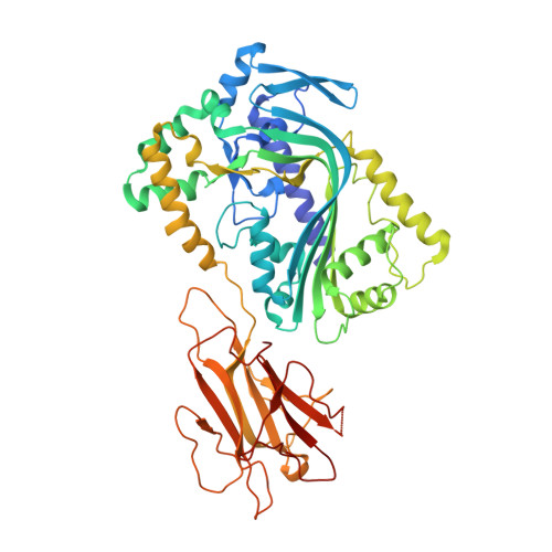
    Proteins containing membrane attack complex/perforin (MACPF) domains play important roles in vertebrate immunity, embryonic development, and neural-cell migration. In vertebrates, the ninth component of complement and perforin form oligomeric pores that lyse bacteria and kill virus-infected cells, respectively. However, the mechanism of MACPF function is unknown. We determined the crystal structure of a bacterial MACPF protein, Plu-MACPF from Photorhabdus luminescens, to 2.0 angstrom resolution. The MACPF domain reveals structural similarity with poreforming cholesterol-dependent cytolysins (CDCs) from Gram-positive bacteria. This suggests that lytic MACPF proteins may use a CDC-like mechanism to form pores and disrupt cell membranes. Sequence similarity between bacterial and vertebrate MACPF domains suggests that the fold of the CDCs, a family of proteins important for bacterial pathogenesis, is probably used by vertebrates for defense against infection.


  • Organizational Affiliation

    Department of Biochemistry and Molecular Biology, Monash University, Clayton, VIC 3800, Australia.


Macromolecules
Find similar proteins by:  (by identity cutoff)  |  3D Structure
Entity ID: 1
MoleculeChains Sequence LengthOrganismDetailsImage
Unknown protein511Photorhabdus laumondii subsp. laumondiiMutation(s): 0 
Gene Names: Photorhabdus luminescens
UniProt
Find proteins for Q7N6X0 (Photorhabdus laumondii subsp. laumondii (strain DSM 15139 / CIP 105565 / TT01))
Explore Q7N6X0 
Go to UniProtKB:  Q7N6X0
Entity Groups  
Sequence Clusters30% Identity50% Identity70% Identity90% Identity95% Identity100% Identity
UniProt GroupQ7N6X0
Sequence Annotations
Expand
  • Reference Sequence
Experimental Data & Validation

Experimental Data

  • Method: X-RAY DIFFRACTION
  • Resolution: 2.00 Å
  • R-Value Free: 0.204 
  • R-Value Work: 0.180 
  • R-Value Observed: 0.182 
  • Space Group: P 21 21 2
Unit Cell:
Length ( Å )Angle ( ˚ )
a = 97.439α = 90
b = 154.507β = 90
c = 43.917γ = 90
Software Package:
Software NamePurpose
DENZOdata reduction
SCALEPACKdata scaling
SHARPphasing
SOLOMONphasing
REFMACrefinement
PDB_EXTRACTdata extraction
HKL-2000data collection

Structure Validation

View Full Validation Report


Entry History 

Revision History  (Full details and data files)

  • Version 1.0: 2007-09-04
    Type: Initial release
  • Version 1.1: 2011-07-13
    Changes: Advisory, Version format compliance
  • Version 1.2: 2017-10-25
    Changes: Refinement description
  • Version 1.3: 2024-03-13
    Changes: Data collection, Database references, Derived calculations