2QLW

Crystal structure of rhamnose mutarotase RhaU of Rhizobium leguminosarum


Experimental Data Snapshot

  • Method: X-RAY DIFFRACTION
  • Resolution: 1.60 Å
  • R-Value Free: 0.180 
  • R-Value Work: 0.152 
  • R-Value Observed: 0.153 

wwPDB Validation   3D Report Full Report


This is version 1.2 of the entry. See complete history


Literature

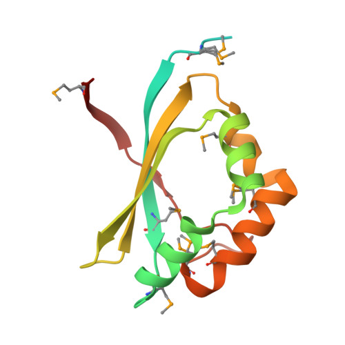
RhaU of Rhizobium leguminosarum is a rhamnose mutarotase.

Richardson, J.S.Carpena, X.Switala, J.Perez-Luque, R.Donald, L.J.Loewen, P.C.Oresnik, I.J.

(2008) J Bacteriol 190: 2903-2910

  • DOI: https://doi.org/10.1128/JB.01120-07
  • Primary Citation of Related Structures:  
    2QLW, 2QLX

  • PubMed Abstract: 

    Of the nine genes comprising the L-rhamnose operon of Rhizobium leguminosarum, rhaU has not been assigned a function. The construction of a Delta rhaU strain revealed a growth phenotype that was slower than that of the wild-type strain, although the ultimate cell yields were equivalent. The transport of L-rhamnose into the cell and the rate of its phosphorylation were unaffected by the mutation. RhaU exhibits weak sequence similarity to the formerly hypothetical protein YiiL of Escherichia coli that has recently been characterized as an L-rhamnose mutarotase. To characterize RhaU further, a His-tagged variant of the protein was prepared and subjected to mass spectrometry analysis, confirming the subunit size and demonstrating its dimeric structure. After crystallization, the structure was refined to a 1.6-A resolution to reveal a dimer in the asymmetric unit with a very similar structure to that of YiiL. Soaking a RhaU crystal with L-rhamnose resulted in the appearance of beta-L-rhamnose in the active site.


  • Organizational Affiliation

    Department of Microbiology, University of Manitoba, Winnipeg, MB R3T 2N2, Canada.


Macromolecules
Find similar proteins by:  (by identity cutoff)  |  3D Structure
Entity ID: 1
MoleculeChains Sequence LengthOrganismDetailsImage
RhaU
A, B
144Rhizobium leguminosarum bv. trifoliiMutation(s): 0 
Gene Names: rhaU
UniProt
Find proteins for Q7BSH1 (Rhizobium leguminosarum bv. trifolii)
Explore Q7BSH1 
Go to UniProtKB:  Q7BSH1
Entity Groups  
Sequence Clusters30% Identity50% Identity70% Identity90% Identity95% Identity100% Identity
UniProt GroupQ7BSH1
Sequence Annotations
Expand
  • Reference Sequence
Small Molecules
Ligands 2 Unique
IDChains Name / Formula / InChI Key2D Diagram3D Interactions
FMT
Query on FMT

Download Ideal Coordinates CCD File 
D [auth A]
E [auth A]
F [auth A]
G [auth A]
H [auth A]
D [auth A],
E [auth A],
F [auth A],
G [auth A],
H [auth A],
I [auth A],
J [auth A],
K [auth A],
L [auth A],
N [auth B],
O [auth B],
P [auth B],
Q [auth B],
R [auth B],
S [auth B],
T [auth B]
FORMIC ACID
C H2 O2
BDAGIHXWWSANSR-UHFFFAOYSA-N
MG
Query on MG

Download Ideal Coordinates CCD File 
C [auth A],
M [auth B]
MAGNESIUM ION
Mg
JLVVSXFLKOJNIY-UHFFFAOYSA-N
Modified Residues  1 Unique
IDChains TypeFormula2D DiagramParent
MSE
Query on MSE
A, B
L-PEPTIDE LINKINGC5 H11 N O2 SeMET
Experimental Data & Validation

Experimental Data

  • Method: X-RAY DIFFRACTION
  • Resolution: 1.60 Å
  • R-Value Free: 0.180 
  • R-Value Work: 0.152 
  • R-Value Observed: 0.153 
  • Space Group: P 32 1 2
Unit Cell:
Length ( Å )Angle ( ˚ )
a = 69.241α = 90
b = 69.241β = 90
c = 101.106γ = 120
Software Package:
Software NamePurpose
DENZOdata reduction
SCALEPACKdata scaling
SHELXphasing
REFMACrefinement
PDB_EXTRACTdata extraction
DNAdata collection
SHELXDphasing

Structure Validation

View Full Validation Report


Entry History 

Deposition Data

Revision History  (Full details and data files)

  • Version 1.0: 2008-11-04
    Type: Initial release
  • Version 1.1: 2011-07-13
    Changes: Advisory, Derived calculations, Version format compliance
  • Version 1.2: 2017-10-18
    Changes: Refinement description