2PVI

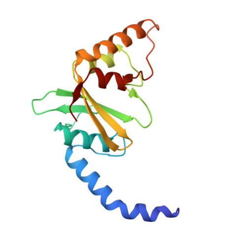
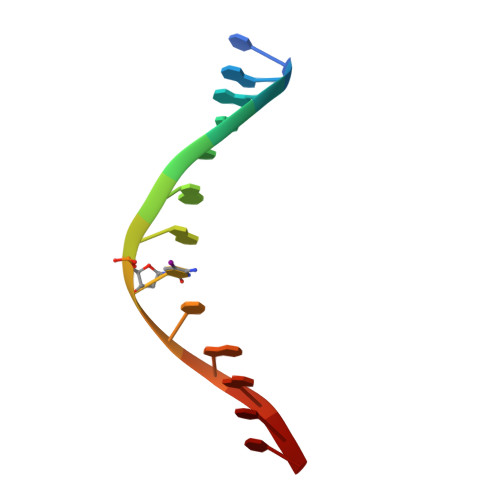
PVUII ENDONUCLEASE COMPLEXED TO AN IODINATED COGNATE DNA


Experimental Data Snapshot

  • Method: X-RAY DIFFRACTION
  • Resolution: 1.76 Å
  • R-Value Free: 0.255 
  • R-Value Work: 0.188 
  • R-Value Observed: 0.188 

wwPDB Validation   3D Report Full Report


This is version 1.4 of the entry. See complete history


Literature

How is modification of the DNA substrate recognized by the PvuII restriction endonuclease?

Horton, J.R.Bonventre, J.Cheng, X.

(1998) J Biol Chem 379: 451-458

  • DOI: https://doi.org/10.1515/bchm.1998.379.4-5.451

  • PubMed Abstract: 

    In restriction-modification systems, cleavage of substrate sites in cellular DNA by the restriction endonuclease is prevented by the action of a cognate methyltransferase that acts on the same substrate sites. The PvuII restriction endonuclease (R.PvuII) has been structurally characterized in a complex with substrate DNA (Cheng et al., 1994) and as an apoenzyme (Athanasiadis et al., 1994). We report here a structure, determined to 1.9 A resolution by crystallography, of a complex between R.PvuII and iodinated DNA. The presence of an iodine at the 5-carbon of the methylatable cytosine results in the following changes in the protein: His84 moved away from the modified base; this movement was amplified in His85 and disrupts an intersubunit hydrogen bond; and the base modification disturbs the distribution of water molecules that associate with these histidine residues and the area of the scissile bond. Considering these observations, hypotheses are given as to why a similar oligonucleotide, where a methyl group resides on the 5-carbon of the methylatable cytosine, is slowly cleaved by R.PvuII (Rice et al., 1995).


  • Organizational Affiliation

    Department of Biochemistry, Emory University School of Medicine, Atlanta, GA 30322, USA.


Macromolecules

Find similar proteins by:  (by identity cutoff)  |  3D Structure
Entity ID: 2
MoleculeChains Sequence LengthOrganismDetailsImage
TYPE II RESTRICTION ENZYME PVUIIC [auth A],
D [auth B]
157Proteus vulgarisMutation(s): 2 
EC: 3.1.21.4
UniProt
Find proteins for P23657 (Proteus hauseri)
Explore P23657 
Go to UniProtKB:  P23657
Entity Groups  
Sequence Clusters30% Identity50% Identity70% Identity90% Identity95% Identity100% Identity
UniProt GroupP23657
Sequence Annotations
Expand
  • Reference Sequence

Find similar nucleic acids by:  Sequence   |   3D Structure  

Entity ID: 1
MoleculeChains LengthOrganismImage
DNA (5'-D(*TP*GP*AP*CP*CP*AP*GP*(C38)P*TP*GP*GP*TP*C)-3')A [auth C],
B [auth D]
13N/A
Sequence Annotations
Expand
  • Reference Sequence
Experimental Data & Validation

Experimental Data

  • Method: X-RAY DIFFRACTION
  • Resolution: 1.76 Å
  • R-Value Free: 0.255 
  • R-Value Work: 0.188 
  • R-Value Observed: 0.188 
  • Space Group: P 21 21 21
Unit Cell:
Length ( Å )Angle ( ˚ )
a = 95.8α = 90
b = 86.3β = 90
c = 48.5γ = 90
Software Package:
Software NamePurpose
X-PLORmodel building
X-PLORrefinement
DENZOdata reduction
SCALEPACKdata scaling
X-PLORphasing

Structure Validation

View Full Validation Report


Entry History 

Deposition Data

Revision History  (Full details and data files)

  • Version 1.0: 1999-12-20
    Type: Initial release
  • Version 1.1: 2007-10-16
    Changes: Version format compliance
  • Version 1.2: 2011-07-13
    Changes: Version format compliance
  • Version 1.3: 2021-11-03
    Changes: Database references, Derived calculations
  • Version 1.4: 2023-08-30
    Changes: Data collection, Refinement description