2PAA

Crystal structure of phosphoglycerate kinase-2 bound to atp and 3pg


Experimental Data Snapshot

  • Method: X-RAY DIFFRACTION
  • Resolution: 2.70 Å
  • R-Value Free: 0.271 
  • R-Value Work: 0.197 

wwPDB Validation   3D Report Full Report


Ligand Structure Quality Assessment 


This is version 1.3 of the entry. See complete history


Literature

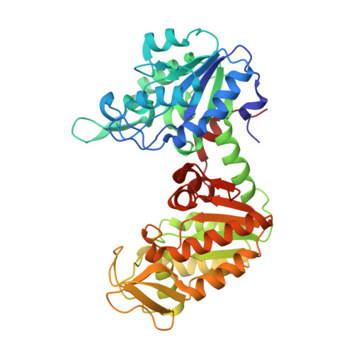
X-ray analysis of phosphoglycerate kinase 2, a sperm-specific isoform from Mus musculus.

Sawyer, G.M.Monzingo, A.F.Poteet, E.C.O'Brien, D.A.Robertus, J.D.

(2007) Proteins 71: 1134-1144

  • DOI: https://doi.org/10.1002/prot.21801
  • Primary Citation of Related Structures:  
    2P9Q, 2P9T, 2PAA

  • PubMed Abstract: 

    Phosphoglycerate kinase 2 (PGK2) is an isozyme of the glycolytic pathway that provides ATP required for sperm motility. It is encoded by an autosomal retrogene that is expressed only during spermatogenesis, concomitant with the inactivation of the X-linked Pgk1 gene. PGK2 from the mouse, Mus musculus, has been overexpressed from a plasmid in bacteria and purified. It was crystallized in three forms: as the apoenzyme, as a complex with 3-phosphoglycerate (3PG), and as a complex with 3PG and ATP. The crystal structures were solved to 2.7, 2.0, and 2.7 A resolutions, respectively. The overall fold is nearly identical with previously solved mammalian PGK1 molecules. The apoenzyme is in the "open" form; that is the N-terminal domain that can bind 3PG and the C-terminal domain that binds ATP are too far apart for the substrates to interact. Binding 3PG causes a 13 degree rotation that partially closes the structure and causes helix 13, which is disordered in the unliganded structure, to stabilize. Binding ATP leaves the protein in the open configuration but also causes helix 13 to be ordered. Sequence alignment suggests that the active site of PGK2 is essentially identical to that of the cytoplasmic PGK1, but significant differences accumulate on a side of the C-terminal domain away from the active site. These changes may mediate the binding of this isoform to other proteins within the sperm flagellum, while still allowing the hinging action between the domains that is essential to catalytic activity.


  • Organizational Affiliation

    Institute for Cellular and Molecular Biology, Department of Chemistry and Biochemistry, University of Texas, Austin TX 78712, USA.


Macromolecules
Find similar proteins by:  (by identity cutoff)  |  3D Structure
Entity ID: 1
MoleculeChains Sequence LengthOrganismDetailsImage
Phosphoglycerate kinase, testis specific
A, B
416Mus musculusMutation(s): 0 
Gene Names: Pgk2Pgk-2
EC: 2.7.2.3
UniProt
Find proteins for P09041 (Mus musculus)
Explore P09041 
Go to UniProtKB:  P09041
Entity Groups  
Sequence Clusters30% Identity50% Identity70% Identity90% Identity95% Identity100% Identity
UniProt GroupP09041
Sequence Annotations
Expand
  • Reference Sequence
Small Molecules
Ligands 2 Unique
IDChains Name / Formula / InChI Key2D Diagram3D Interactions
ATP
Query on ATP

Download Ideal Coordinates CCD File 
C [auth A]ADENOSINE-5'-TRIPHOSPHATE
C10 H16 N5 O13 P3
ZKHQWZAMYRWXGA-KQYNXXCUSA-N
3PG
Query on 3PG

Download Ideal Coordinates CCD File 
D [auth B]3-PHOSPHOGLYCERIC ACID
C3 H7 O7 P
OSJPPGNTCRNQQC-UWTATZPHSA-N
Binding Affinity Annotations 
IDSourceBinding Affinity
3PG BindingDB:  2PAA Kd: 1.09e+5 (nM) from 1 assay(s)
Experimental Data & Validation

Experimental Data

  • Method: X-RAY DIFFRACTION
  • Resolution: 2.70 Å
  • R-Value Free: 0.271 
  • R-Value Work: 0.197 
  • Space Group: P 1
Unit Cell:
Length ( Å )Angle ( ˚ )
a = 39.854α = 90.34
b = 64.541β = 77.2
c = 75.936γ = 85.03
Software Package:
Software NamePurpose
DENZOdata reduction
SCALEPACKdata scaling
CNSrefinement
PDB_EXTRACTdata extraction
CrystalCleardata collection
HKL-2000data reduction
HKL-2000data scaling
MOLREPphasing

Structure Validation

View Full Validation Report


Ligand Structure Quality Assessment 


Entry History 

Deposition Data

Revision History  (Full details and data files)

  • Version 1.0: 2007-11-27
    Type: Initial release
  • Version 1.1: 2011-07-13
    Changes: Version format compliance
  • Version 1.2: 2017-10-18
    Changes: Refinement description
  • Version 1.3: 2023-08-30
    Changes: Data collection, Database references, Derived calculations, Refinement description