2P28

Structure of the PHE2 and PHE3 fragments of the integrin beta2 subunit


Experimental Data Snapshot

  • Method: X-RAY DIFFRACTION
  • Resolution: 2.20 Å
  • R-Value Free: 0.308 
  • R-Value Work: 0.261 
  • R-Value Observed: 0.263 

wwPDB Validation   3D Report Full Report


This is version 1.3 of the entry. See complete history

Re-refinement Note

A newer entry is available that reflects an alternative modeling of the original data: 5E6W


Literature

A structural hypothesis for the transition between bent and extended conformations of the leukocyte beta2 integrins

Shi, M.Foo, S.Y.Tan, S.M.Mitchell, E.P.Law, S.K.A.Lescar, J.

(2007) J Biol Chem 282: 30198-30206

  • DOI: https://doi.org/10.1074/jbc.M701670200
  • Primary Citation of Related Structures:  
    2P26, 2P28

  • PubMed Abstract: 

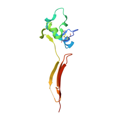
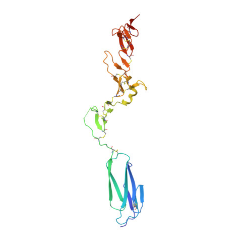
    Integrins mediate cell adhesion in response to activation signals that trigger conformational changes within their ectodomain. It is thought that a compact bent conformation of the molecule represents its physiological low affinity state and extended conformations its active state. We have determined the structure of two integrin fragments of the beta2 subunit. The first structure, consisting of the plexin-semaphorin-integrin domain, hybrid, integrin-epidermal growth factor 1 (I-EGF1), and I-EGF2 domains (PHE2), showed an L-shaped conformation with the bend located between the I-EGF1 and I-EGF2 domains. The second structure, which includes, in addition, the I-EGF3 domain, showed an extended conformation. The major reorientation of I-EGF2 with respect to the other domains in the two structures is accompanied by a change of torsion angle of the disulfide bond between Cys(461)-Cys(492) by 180 degrees and the conversion of a short alpha-helix (residues Ser(468)-Cys(475)) into a flexible coil. Based on the PHE2 structure, we introduced a disulfide bond between the plexin-semaphorin-integrin domain and I-EGF2 domains in the beta2 subunit. The resultant alphaLbeta2 integrin (leukocyte function-associated antigen-1) variant was locked in a bent state and could not be detected with the monoclonal antibody KIM127 in Mg(2+)/EGTA. However, it retained the binding activity to ICAM-1. These results provide a structural hypothesis for our understanding of the transition between the resting and active states of leukocyte function-associated antigen-1.


  • Organizational Affiliation

    School of Biological Sciences, Nanyang Technological University, 60 Nanyang Drive, Singapore, Singapore 637551.


Macromolecules
Find similar proteins by:  (by identity cutoff)  |  3D Structure
Entity ID: 1
MoleculeChains Sequence LengthOrganismDetailsImage
Integrin beta-2100Homo sapiensMutation(s): 0 
UniProt & NIH Common Fund Data Resources
Find proteins for P05107 (Homo sapiens)
Explore P05107 
Go to UniProtKB:  P05107
PHAROS:  P05107
GTEx:  ENSG00000160255 
Entity Groups  
Sequence Clusters30% Identity50% Identity70% Identity90% Identity95% Identity100% Identity
UniProt GroupP05107
Sequence Annotations
Expand
  • Reference Sequence
Find similar proteins by:  (by identity cutoff)  |  3D Structure
Entity ID: 2
MoleculeChains Sequence LengthOrganismDetailsImage
Integrin beta-2217Homo sapiensMutation(s): 0 
UniProt & NIH Common Fund Data Resources
Find proteins for P05107 (Homo sapiens)
Explore P05107 
Go to UniProtKB:  P05107
PHAROS:  P05107
GTEx:  ENSG00000160255 
Entity Groups  
Sequence Clusters30% Identity50% Identity70% Identity90% Identity95% Identity100% Identity
UniProt GroupP05107
Sequence Annotations
Expand
  • Reference Sequence
Small Molecules
Ligands 1 Unique
IDChains Name / Formula / InChI Key2D Diagram3D Interactions
NAG
Query on NAG

Download Ideal Coordinates CCD File 
C [auth A]2-acetamido-2-deoxy-beta-D-glucopyranose
C8 H15 N O6
OVRNDRQMDRJTHS-FMDGEEDCSA-N
Experimental Data & Validation

Experimental Data

  • Method: X-RAY DIFFRACTION
  • Resolution: 2.20 Å
  • R-Value Free: 0.308 
  • R-Value Work: 0.261 
  • R-Value Observed: 0.263 
  • Space Group: P 65 2 2
Unit Cell:
Length ( Å )Angle ( ˚ )
a = 52.26α = 90
b = 52.26β = 90
c = 423.924γ = 120
Software Package:
Software NamePurpose
AMoREphasing
REFMACrefinement
PDB_EXTRACTdata extraction
MOSFLMdata reduction
SCALAdata scaling
PHASESphasing

Structure Validation

View Full Validation Report


Entry History 

Deposition Data

Revision History  (Full details and data files)

  • Version 1.0: 2007-08-14
    Type: Initial release
  • Version 1.1: 2011-07-13
    Changes: Non-polymer description, Version format compliance
  • Version 1.2: 2020-07-29
    Type: Remediation
    Reason: Carbohydrate remediation
    Changes: Advisory, Data collection, Database references, Derived calculations, Structure summary
  • Version 1.3: 2023-10-25
    Changes: Data collection, Database references, Refinement description, Structure summary