


Anion-dependent hinge motion in ferric binding proteins
Tom-Yew, S.A.L., Shilton, B.H., Bekker, E.G., Tocheva, E.I., Murphy, M.E.P.To be published.
Experimental Data Snapshot
wwPDB Validation 3D Report Full Report

Entity ID: 1 | |||||
|---|---|---|---|---|---|
| Molecule | Chains | Sequence Length | Organism | Details | Image |
| Putative iron binding protein | 323 | Bordetella pertussis Tohama I | Mutation(s): 0 Gene Names: bFbpA | 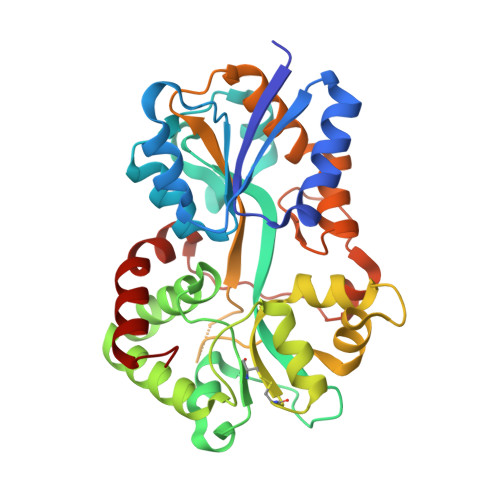 | |
UniProt | |||||
Find proteins for Q7VXW9 (Bordetella pertussis (strain Tohama I / ATCC BAA-589 / NCTC 13251)) Explore Q7VXW9 Go to UniProtKB: Q7VXW9 | |||||
Entity Groups | |||||
| Sequence Clusters | 30% Identity50% Identity70% Identity90% Identity95% Identity100% Identity | ||||
| UniProt Group | Q7VXW9 | ||||
Sequence AnnotationsExpand | |||||
| |||||
| Ligands 2 Unique | |||||
|---|---|---|---|---|---|
| ID | Chains | Name / Formula / InChI Key | 2D Diagram | 3D Interactions | |
| CO3 Query on CO3 | C [auth A] | CARBONATE ION C O3 BVKZGUZCCUSVTD-UHFFFAOYSA-L |  | ||
| FE Query on FE | B [auth A] | FE (III) ION Fe VTLYFUHAOXGGBS-UHFFFAOYSA-N |  | ||
| Length ( Å ) | Angle ( ˚ ) |
|---|---|
| a = 54.674 | α = 90 |
| b = 57.278 | β = 90 |
| c = 87.927 | γ = 90 |
| Software Name | Purpose |
|---|---|
| REFMAC | refinement |
| HKL-2000 | data collection |
| HKL-2000 | data reduction |
| HKL-2000 | data scaling |
| AMoRE | phasing |
RCSB PDB (citation) is hosted by
RCSB PDB is a member of the

